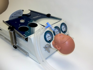
Grand Rapids, MI – Grand Valley State University students will soon have the opportunity to simulate the proper management of life-like respiratory ailments using the latest in training and test lung devices. Grand Rapids-based, Michigan Instruments Inc. developers of the world-renowned “Michigan Lung” plans to donate two TTL respiratory simulation units to the program, with a value of approximately $25,000. Grand Valley and Muskegon Community Colleges are collaborating to offer Muskegon’s Respiratory Therapy education for GVSU students. Nursing and Physician Assistant students at GVSU will also benefit from the simulation units.

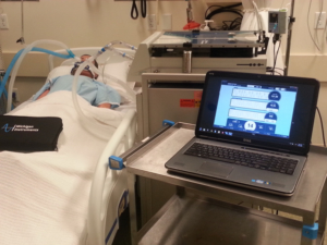
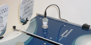
These sophisticated devices provide students with real-time data, measurements, and response that simulate those of a real respiratory patient. With this information, students learn how to properly ventilate and manage a variety of respiratory conditions.

“It is our privilege to provide the latest advancements in training and test lung products to a local program like Grand Valley State University’s,” stated Joe Baldwin, President of Michigan Instruments, Inc. “Our ‘Test Lungs’ are known and recognized worldwide and we are fortunate to work with a program like GVSU’s to ensure students in our community are able to receive the absolute best training possible right here in West Michigan.”

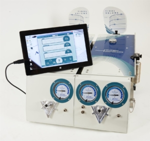
Michigan Instruments Inc, partnered with local software design and development firm, Atomic Object, to architect and develop cutting-edge software called “PneuView 3” — their latest training and test lung software application which calculates and displays, in real time, numerous respiratory parameters and waveforms. Software improvements combined with intricate design modifications to the Michigan Lung, provide users with even greater simulation capabilities.

The Michigan Lung is regarded as the most versatile, reliable training and test lungs on the market, and its latest multifaceted, fully to-scale mechanical design and software upgrades allow for simulation of hundreds of patient scenarios.

About Michigan Instruments

MII has designed and manufactured specialized medical equipment related to the fields of cardiovascular medicine, mechanical CPR, and respiratory therapy for over forty years. The company has built a reputation for medical device products of exceptional quality, which has earned the respect of thousands of customers, associates, and medical professionals throughout the world.

MII complies with Food and Drug Administration (FDA) and International Organization of Standardization (ISO) regulations for Good Manufacturing Practices. Both the FDA and ISO systems require continuous control over all activities that assure the quality of Michigan Instruments products and services. MII has built a strong foundation for growth based on the dedication of its’ staff, a close relationship For more information, contact us today.

Michigan Instruments would like to announce a change in cost associated with the calibration of many Training and Test Lung respiratory simulation models.

On November 1, 2014, the cost for models 1600, 1601, 1603 and 4600 (recommended every 3 years) and models DAN, AIN and SLN (recommended every 2 years) will be $560. For models 3600i, 5600i, 5601i, DA3, AI3, and SL3 (recommended every 2 years) will be $700.

There will no longer be a separate for charge for “as found” or “as received” data. This data will be taken (if the unit is received in a condition to permit it) and provided with final data and certifications.

Factory calibration encompasses all aspects of sensor, electronic, mechanical and software tuning to ensure maximum accuracy of all measurements and simulated parameters provided by TTL/PneuView systems. This procedure includes the setting of the compliance and resistance characteristics of the lungs and setting offset and gain characteristics for each of the pressure transducer channels.

Is it possible to do this independently?

Pressure and volume calculation accuracy may be easily verified with the aid of a calibrated syringe and independent pressure measurement device. All versions of the PneuView system ship with pre-formatted templates to aid the user with the verification of calibration accuracy. These templates include step-by-step instructions, which should be followed periodically to ensure maintenance of calibration accuracy or anytime accuracy comes into question, such as if the unit suffers some type of physical damage.

To schedule an appointment with Michigan Instruments click here.

To contact our Service Department:

Phone: 800-530-9939
Extension: 343
E-Mail: service@michinst.com

It’s no secret—mechanical systems move differently than biological systems particularly when it comes to inertia. This concept has a number of significant implications when working with a mechanical test lung.

During ventilation, the inertia of the lung chamber must first be overcome before it can start to expand. Prior to this point, any gas delivered to the lung has been delivered into a chamber with static compliance. Only when the lung chamber begins to move do we see the dynamic compliance changes that would be expected of a normal patient. This results in a sharp peak in pressure data taken at the beginning of inspiration as the lung chamber overcomes inertia and at the end of expiration as the chamber returns to rest. Static compliance compensation is rarely included in ventilators and other medical instruments since it does not represent a normal patient scenario.

Physical Symptoms of Inertia

The most common physical symptom of this problem occurs during the expiratory phase. When a test lung with considerable inertia returns to rest at the end of a breath it can cause a pressure “bounce” in the airway that is sometimes strong enough to trigger an assisted breath from an IMV/SIMV compatible machine. While the false peaks in the pressure wave are rarely large enough to trigger limit warnings in a ventilator, this second breath caused by the bounce can skew tests measuring rate parameters such as breath rate and minute volume.

How to Resolve Measuring Rate Parameters

While frustrating, this problem is not unsolvable. Some mechanical test lungs are equipped with counterbalances to help minimize the inertia of the lung chamber while advanced breath parsing in instrumentation allows knowledgeable users to eliminate most of the false peaks that come up in testing. Tests themselves are often adjusted to get rid of these inaccuracies.

Almost all modern ventilation tests call for a PEEP of at least 5 cmH2O which (while being representative of actual ventilation practices) helps to reduce the inertia of mechanical and biological lungs alike. Decreasing I:E ratio can also help as it allows for a more gradual progression between the breath phases.

If you have any questions regarding the role inertia plays in mechanical test lungs, feel free to contact Michigan Instruments anytime, we are here to help!

A number of our customers have been requesting information on how to simulate a pneumothorax using the Michigan Instruments Dual Adult Lung. The following will describe how this condition can be simulated on a Training & Test Lung, using setup options offered by this device. The principles described can be applied to many laboratory situations that do not require the use of this lung.

A pneumothorax is usually characterized in humans by a collection of gas or fluid between a lung and the wall of the chest cavity. The added pressure (usually on only one lung) created by this condition often results in a patient having one uncompliant lung and one healthy lung—a situation in which standard ventilation can be difficult or even dangerous to perform.

The Simulation

This scenario can be simulated on a model 1600 or 5600i TTL by reducing the compliance of one lung (usually to 0.02 or 0.03 liters/cmH2O) while leaving the second lung in a healthy state (0.05 liters/cmH­2O).

Airway resistances should be left within the normal healthy range (Rp20 for upper airway, Rp5 for lower airway) unless a compound pneumothorax is being simulated. When a TTL set up in this manner is ventilated the operator will note that, while pressures are relatively even throughout the system, volume and flow are heavily skewed toward one lung (as it would be in an actual patient scenario).

The pneumothorax can be accentuated (for observational purposes only) by further reducing the compliance of the affected lung (to 0.01 liters/cmH2O) and setting the upper and both lower airway resistances to Rp20. This extreme scenario, while effective at conveying the effects of a pneumothorax, is not representative of a simple pneumothorax in humans and should not be used to assess the effectiveness of treatment methods for this condition.

Most test lungs can be used to simulate many different pulmonary conditions. If you have questions on how to create a specific scenario on your test lung please leave a comment below or contact techsupport@michinst.com. More information is available at https://www.michiganinstruments.com/training-test-lungs/.

It is no secret that military spending has gone down since sequestration (and other spending cuts), and simulation was one of the areas to take a major hit from this. This has led to increased private simulation lab interest and funding.

I remember attending the International Meeting on Simulation in Healthcare at the San Diego Convention Center in 2012. The sounds of my automated ventilator were constantly drowned out by faux gunfire and helicopter blades. Half of the people that walked by my booth were in uniform. Mannequins sported ragged gashes, shrapnel and even face paint.

Fast forward to 2014 in Orlando, these curiosities were nowhere to be found. Instead many of the rubber cadavers wore hospital gowns. Plenty of grave injuries made themselves present, but they were along the lines of blocked airways and organ complications. The focus of this industry is changing. While military spending is down, private spending in the realm of simulation is still very high. Simulation labs are becoming a new standard in modern hospitals, and those on the cutting edge of this industry are always looking for a way to improve their users’ experience.

Private User Attractiveness

As this change occurs, companies are beginning to ask questions about what will make their devices more appealing to private users. Cost is clearly not an issue. In 2012, a very high-end lung simulator would have cost between 25,000 and 35,000 USD. Just last week, at a show in Paris, I was introduced to a new lung simulation system with a base price of 46,000 Euros—well over 60,000 USD at the time.

Key Factors in Increasing Private Lab Simulation Attention

So, if cost is not an issue, how do we tap into this growing wealth of enthusiasm for simulation equipment? I won’t claim to have all of the answers, but customers have repeatedly asked questions that lead me to believe that the key factors are:

  1. Anthropomorphism—Simulation lab directors tend to want their hardware to look human. This helps users to relate to the procedures that they are performing and increases the learning experience.
  2. Durability—No one wants to spend 60,000 dollars on a device that will not last. Some of the most marketable simulation devices that I have seen have boxy steel frames or rugged, built-in carts that make transportation safe and easy.
  3. Availability of Support—It’s a lot easier to be confident buying a unit with a substantial guarantee, warranty, or support policy.
  4. The Range of Use—Very few customers will buy an extravagant device to simulate a single condition. The most successful simulators have a wide range of both “normal” and “abnormal” settings.
  5. Reputation—The industry talks. If one intubation simulator has been bought by the majority of the simulation labs in a given area, new simulation labs nearby are likely to purchase it. It is becoming more important than ever to maintain excellent relationships with customers, as both complaints and praise can be spread worldwide on popular media sites.

The industry is in a constant state of flux of change; at Michigan Instruments, we always keep our ear to the ground and function in a consistent state of improvement, ensuring quality products no matter what. Reach out to us anytime to learn more and see how we can best help you.

Acute Respiratory Distress Syndrome, or ARDS, is by no means a new condition in the respiratory care industry, but there are certainly some new treatments being tested to help treat it. Herneations, H1N1, SARS and many other conditions are all forms of ARDS, and it can affect patient of almost any age. High-frequency oscillatory ventilation (or HFOV) is an innovative form of respiratory care that, in America, is not widely used or understood. Michigan Instruments provides an explanation below.

High-frequency oscillatory ventilation explained:

Fortunately, the concept is simple. A healthy person in need of respiratory assistance is usually ventilated at a rate of 12-14 breaths per minute, each consisting of 400-700mL of air. HFOV, on the other hand, will deliver tiny ventilations, perhaps about 10-30mL, hundreds of times every minute. These tiny injections are understood to put less strain on the already distressed lungs than the conventional 600mL ventilations.

Unfortunately there are very few testers that can analyze the performance of an HFOV ventilator, and it is for this reason, at least in part, that the practice has not become more widespread. Flow measurements are particularly hard to perform on HFOV injections, and the diffusion path of delivered gas through the respiratory system is almost impossible to monitor.

In part two of this 3-part series, Michigan Instruments will dive into the details regarding output assessment and how to properly monitor the effectiveness of this technique.

If you have any questions about the use of high-frequency oscillatory ventilation, don’t hesitate to contact Michigan Instruments anytime.

This is part 2 of our series focusing on assessing high-frequency oscillatory ventilation. Check out part one here.

Due to difficulties in assessing the outputs (from the patient’s perspective) of HFOV ventilators, many analysts have resorted to monitoring pressure outputs. The theory is that so long as an adequate FIO2 (fraction of inspired oxygen) reaches the patient and the pressures remain safe, the ventilation is a success. Unfortunately, this method does little to help us understand HFOV on a deeper level, and without this understanding, it is difficult to know how the science and practice of modern ventilation will progress in the future.

Why is it important to understand volume and flow parameters?

Measuring ventilated volume is a common way of fact-checking a ventilator. For the display on a ventilator to show an accurate delivered volume the vast majority of the unit’s internal systems must be working properly. Flow, similarly, can offer insight into what might be going wrong with one of these ventilators, should it malfunction or require calibration. These parameters are sometimes overlooked in testing as the ventilator itself will display variations of them. Other variables make themselves in apparent when the problem of calibration is approached scientifically. Compressible volume in the airway and lungs can damage the accuracy of calculations performed by the ventilator, and without the ability to control airway resistance and lung compliance to a known value outputs are not always consistent.

The patient’s perspective.

Changes in flow and volume must be explored from the perspective of the patient as a way to understand, calibrate, and tweak ventilators. This sort of testing, however, requires a new type of test lung device capable of measuring flows, volumes, and pressures at different points throughout the respiratory system of a simulated patient.

We’ll get into test lung devices in part three. In the meantime, if you have any questions about the use of high-frequency oscillatory ventilation, don’t hesitate to contact Michigan Instruments anytime.

In using high frequency oscillatory ventilation (HFOV) tidal volume and LPM (flow) values can be exceptionally difficult to monitor. Several third-party devices have been developed to monitor this, but due to a lack of any solid standard of operation, it is still considered to increase patient risk over conventional ventilation. Regardless, let’s take a look at its current applications and uses.

Currently, HFOV has found a niche in non-conventional settings, primarily in the treatments of neonatal patients and some adult cases where the patient is considered to be past the point at which traditional ventilation would be beneficial. It is important to note that the largest fear surrounding HFOV concerns is the lack of understanding of the delivered tidal volume and other common parameters used to describe breaths.

HFOV, in some medical systems, has become a “last resort” treatment option.

The Benefit

It doesn’t expose the lungs of a patient to the same dramatic swings in pressure and flow that a standard ventilator does. Damaged lungs can be further exacerbated by such swings, and the underdeveloped lungs of premature infants are especially susceptible.

The Drawbacks

HFOV is such a departure from a standard breath that it invites mistrust, and without the ability to really explore the WHY of the effectiveness of HFOV, such a departure is difficult to justify. It is often found, in medical studies, that the human body is very well adapted to its’ natural processes.

The mention of a ventilator forcing hundreds of miniature breaths onto a patient every minute is, admittedly, a bit intimidating. It could, however, offer some excellent treatment options to future patients. We aren’t saying advocating to press this sort of treatment—not until we find a way to confirm effectiveness and minimize risk to patients—but we do believe that the benefits of better understanding this non-conventional form of ventilation would far outweigh the cost.

Review part one and part two of the series and contact us with any follow-up questions or comments.