Lung simulation is essential for healthcare professionals to practice respiratory care skills in a safe, controlled environment. However, even with the best equipment, challenges can arise that can disrupt training…

When you hear the word “test lung” and “lung simulator,” you may think they mean the same thing. Many people use them simultaneously. In fact, we have historically called our lung simulators “Training & Test Lungs “or TTLs – since they have several lung simulation, teaching and testing…

what-to-do-in-a-cardiac-emergency

Cardiac emergencies can strike suddenly and without warning. Whether it’s a heart attack or a cardiac arrest, knowing how to respond in these crucial moments can save a life. 

In this blog, we’ll walk you through a step-by-step guide on what to do during a cardiac emergency, emphasizing quick actions and the importance of reliable medical equipment to assist in these life-threatening situations.

Step 1: Assess the Situation

When faced with a potential cardiac emergency, the first step is to assess the situation. Check for signs of cardiac arrest, such as unresponsiveness and abnormal and/or no breathing. 

If the person is unresponsive and not breathing, this may indicate a cardiac arrest, which requires immediate action.

Step 2: Call for Help

Dial 911 immediately. This ensures that emergency services are on their way and can provide advanced care as quickly as possible. If you are in a healthcare setting, alert other medical professionals to the situation.

Step 3: Start Chest Compressions

Chest compressions are  a critical first step in saving a life during a cardiac arrest. Place the heel of your hand on the center of the chest, just below the sternum. 

Use your other hand to support it and begin compressing the chest at least 2 inches deep at a rate of 100-120 compressions per minute. Be sure to allow full chest recoil after each compression.

Step 4: Administer Defibrillation (If Available)

If an automated external defibrillator (AED) is nearby, turn it on immediately and follow the instructions provided. The AED is programmed to  analyze the person’s heart rhythm and, if necessary, deliver a shock to restore normal heart activity. Continue CPR until the AED is ready to administer a shock or until emergency personnel arrive.

Step 5: Continue CPR

If an AED is not available, continue CPR until help arrives. If you are trained in advanced techniques, you may also consider performing rescue breaths, especially in cases where the person is a child or an infant. 

For untrained responders, hands-only CPR (chest compressions without breaths) is still highly effective in improving survival outcomes.

Step 6: Provide Post-Resuscitation Care

Once emergency responders arrive, they will take over the care and transport the individual to the hospital for further treatment. Follow any additional instructions given by medical professionals, and ensure you remain calm throughout the process.

How Michigan Instruments Helps in Cardiac Emergencies

Cardiac emergencies demand rapid and effective interventions, and the right tools can significantly impact the outcomes. Michigan Instruments has been a leader in life-saving technology since 1964, supporting healthcare professionals and first responders with reliable and easy-to-use solutions.

Our automated CPR devices are designed to deliver precise and consistent compressions, allowing medical teams to focus on what matters most — saving lives. Whether in the field or at the hospital, Michigan Instruments empowers those on the front lines to provide optimal care in critical moments.

With over 60 years of experience, we remain committed to improving patient outcomes and supporting the heroes who respond to cardiac emergencies.

Learn more about how our advanced automated CPR devices can make a difference for your team.

The Anatomy of a Lung Simulator How It Works

Lung simulators play a crucial role in respiratory therapy, critical care training, medical research and setting a standard for replicating human lung function in controlled settings. By mimicking human lung function, lung simulators offer healthcare professionals and researchers a reliable tool to improve patient care and deepen the understanding of respiratory mechanics. 

In this blog, we’ll break down some of the components and functions of lung simulators, highlighting their innovative design and practical applications.

What are Lung Simulators?

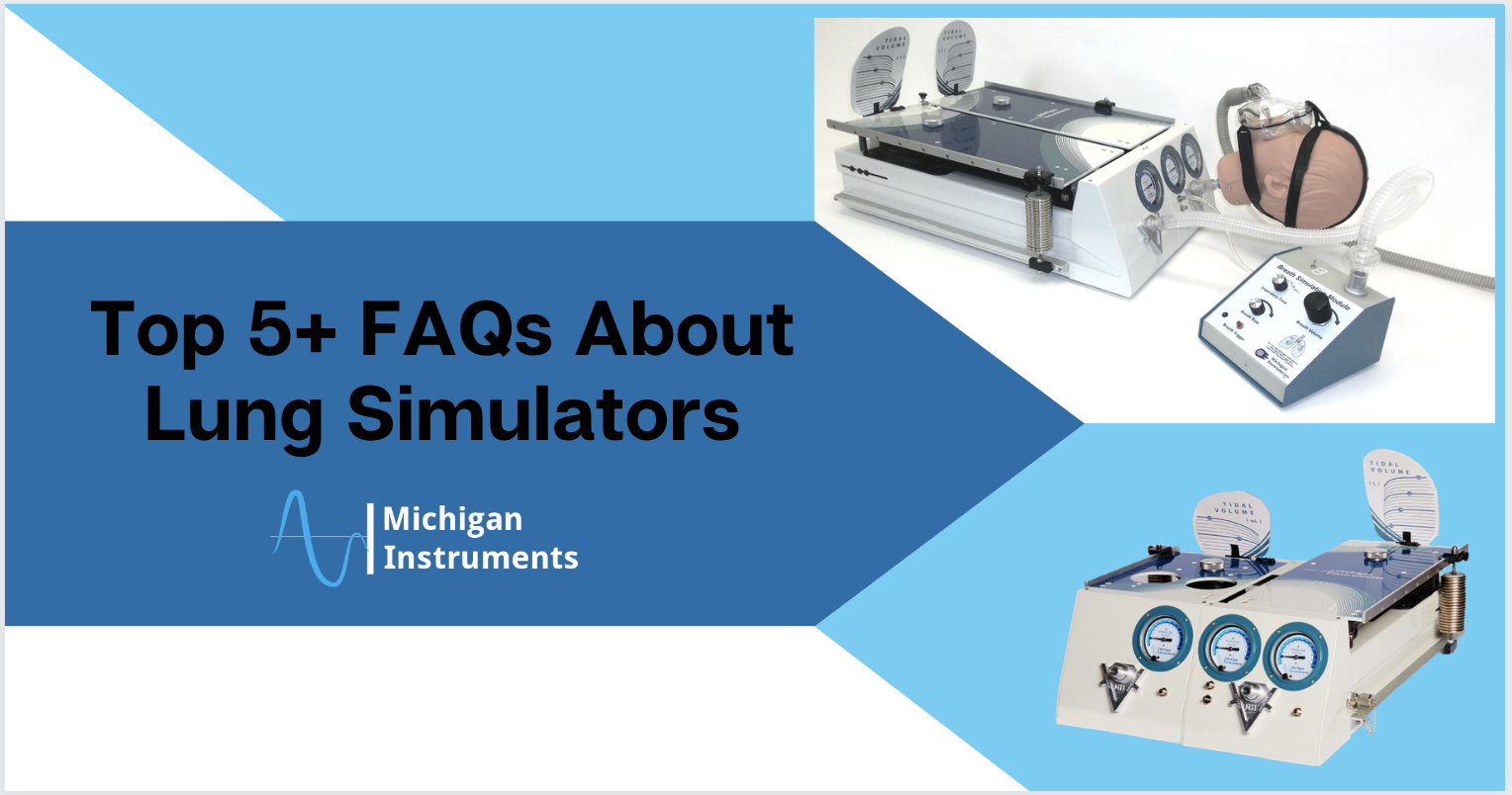
There are several variations available, but overall, a lung simulator is a precision instrument designed to replicate the mechanical properties of human lungs. They allow users to simulate various breathing conditions, lung capacities, and respiratory challenges.

Lung simulators are invaluable for teaching and training, testing ventilators, and conducting clinical research. They offer a safe and controlled environment to explore complex respiratory scenarios.

Key Components of Lung Simulators

1. Test Lung Chambers

The test lung chambers form the centerpiece of a lung simulator. These chambers are designed and set up to mimic the elasticity and compliance of human lungs. 

Most have the capability to replicate various lung conditions by offering settings from healthy lung function to diseases such as Chronic Obstructive Pulmonary Disease (COPD) or Acute Respiratory Distress Syndrome (ARDS).

2. Resistance Elements

Resistance elements control airflow within the simulator, replicating the resistance encountered in a patient’s airways. By adjusting the resistance, users can simulate different airway conditions. This offers versatility in testing ventilators or teaching/training in practical clinical scenarios.

3. Pressure and Volume Sensors

There are sensors embedded into some simulators that measure pressure, volume, and airflow in real time. These sensors provide critical data that help the user evaluate device performance, patient safety, and the efficacy of various treatment strategies.

4. External Connections

Lung simulators are designed with  connections for ventilators, oxygen delivery systems, and other respiratory devices. These connections allow the simulator to serve as a testing platform, ensuring the performance and safety of life-supporting equipment.

How Does a Lung Simulator Work?

The lung simulator is designed to replicate the dynamics of human respiration. Below is an overview of how that’s done: 

  1. Setup: The simulator is configured to mimic the desired respiratory condition. This may involve adjusting lung compliance, airway resistance, or the breathing pattern.
  2. Simulation: Airflow is introduced to the test lung chambers through external devices such as ventilators. The simulator responds by producing a realistic response and lung mechanics in the same way a patient would.
  3. Measurement: Data such as pressure, volume, and flow are collected, providing valuable insights into device performance and patient conditions.
  4. Analysis: The collected data is analyzed and the users are able to evaluate the effectiveness of respiratory therapies, devices, or clinical interventions.

Applications of Lung Simulators

Lung simulators are used across a wide range of fields, including:

  • Medical Training: Providing hands-on experience for healthcare professionals to practice managing respiratory conditions and operating ventilators.
  • Device Testing: Ensuring ventilators and other respiratory devices perform effectively under various conditions.
  • Research: Facilitating studies into respiratory diseases and the development of new treatment methods.
  • Education: Demonstrating respiratory mechanics to students and trainees in medical and healthcare programs.

Why Choose Michigan Instruments for Lung Simulators?

For decades, Michigan Instruments has led the way in lung simulator innovation, providing industry-leading tools for medical research and training. Our devices are known for their precision, reliability, and versatility. Whether you’re testing life-saving equipment or advancing medical research, our lung simulators provide the tools you need to succeed.

Explore the features and benefits of our lung simulators today. With Michigan Instruments, you’re not just getting a product – you’re gaining a partner in innovation and excellence.

For more insights and updates on medical simulation technology, follow our blog or contact us today. Let’s work together to shape the future of respiratory care.

respiratory therapy for veterans

At Michigan Instruments, we believe that Veterans are a crucial part  of our nation. They’ve made incredible sacrifices to fight for our freedom, and they deserve exceptional care– not only during their service but throughout their lives. Some of the conditions they face during their time in service may lead to unique healthcare challenges, including respiratory issues that require specialized attention. 

In fact, studies show that over 12% of veterans have reported some kind of obstructive respiratory disease. Offering these heroes the best possible respiratory care, including access to advanced lung simulators, is a critical mission in itself. Providing high-quality respiratory therapy for veterans is not just a necessity—it is a vital step toward enhancing their quality of life. Thanks to advancements in medical technology, VA clinics are better equipped than ever to address these needs.

The Importance of Respiratory Therapy for Veterans

Veterans often experience respiratory issues linked to their time in service, such as:

Diagnosing lung and breathing conditions is the first step to determining what type of respiratory therapy for veterans is needed to help improve lung function and overall health. VA clinics are tasked with addressing these issues and leveraging innovative solutions to provide targeted care for their veteran patients.

How Advanced Lung Simulators Improve Veteran Care

Lung simulators are high-tech devices that replicate human lung function, offering a range of benefits for respiratory therapy training, research, and patient care. Here’s how they enhance care in VA clinics across the country, such as the CARE Comprehensive Assessment of Respiratory and other military Exposures Center:

1. Improved Training for Respiratory Therapists

While VA clinics rely on well-trained respiratory therapists to deliver specialized care, advanced lung simulators further help to provide a low-risk training environment by offering:

  • Realistic Scenarios: Simulators mimic various lung conditions, enabling therapists to practice diagnosing and treating diverse respiratory challenges.
  • Hands-On Experience: Trainees can work on mastering mechanical ventilation and other life-saving techniques in a risk-free environment.
  • Confidence Building: Simulators help therapists gain confidence and competence before working directly with patients, including the Nation’s finest.

2. Enhanced Research Capabilities

Research is a huge part of advancing respiratory therapy for veterans. Many studies on respiratory health have been conducted by VA research teams focused on covering a wide range of topics, including tuberculosis, lung cancer and smoking, influenza, and pneumonia. Within VA research facilities, lung simulators allow researchers to:

  • Test New Ventilation Techniques by evaluating the effectiveness of emerging therapies under controlled conditions.
  • Study Chronic Conditions by investigating the progression of diseases like COPD and asthma to develop better treatment plans.
  • Simulate Real-World Scenarios by exploring how respiratory conditions interact with various environmental factors, such as high altitudes or exposure to toxins.

3. Better Patient Outcomes

By integrating advanced lung simulators into care protocols, VA clinics can:

  • Personalize Treatment Plans using data-driven insights from simulators to tailor respiratory therapy to each veteran’s needs.
  • Improve Ventilator Settings by optimizing ventilator configurations to ensure effective and comfortable breathing support.
  • Advance Emergency Preparedness by equipping teams to respond to acute respiratory crises with greater precision and speed.

Michigan Instruments is Ready for Service as Your Partner in Veteran Care

Michigan Instruments has long been a leader in developing cutting-edge lung simulation technology. In fact, we’ve been at it since the 1960’s, when the US got involved in the Vietnam War! Our products are designed to empower healthcare professionals with the tools they need to provide exceptional care for patients of all types. Whether training new therapists or enhancing research, our lung simulators play a vital role in improving respiratory therapy for veterans.

Medical facilities across the country are turning to Michigan Instruments’ simulators for training and research. By investing in these advanced tools, we can ensure that veterans receive the care they deserve.

Fight Respiratory Disease with Michigan Instruments 

As we continue to prioritize veteran care and advancing respiratory therapy for veterans, Michigan Instruments remains dedicated to supporting healthcare providers with the technology and expertise needed to make a difference. By using tools like advanced lung simulators, VA clinics are better equipped to meet the unique respiratory health challenges veterans face.

rare respiratory diseases

In the United States, a rare disease is defined by a prevalence of < 200,000 patients. There are approximately 7,000 unique rare diseases affecting an estimated 25 to 30 million people in the United States. So researching these conditions is incredibly important.

About 5-10% of rare disease diagnoses are lung-related, such as Alpha-1 antitrypsin deficiency, Pulmonary arterial hypertension, and Lymphangioleiomyomatosis (LAM). These conditions may be foreign to most, but to the people who suffer from them and the researchers who study them, they are all too familiar. 

Restoring Hope for Rare Respiratory Disease Researchers

Rare respiratory disease can cause  barriers in a patients life, and the rarity of their disease can be isolating. Unfortunately, researchers of rare respiratory disease can feel isolated as well; With only a few cases, clinical data and research opportunities are limited in both clinical research and treatment development.

Luckily, advanced lung simulators have transformed how we study rare diseases, making it possible for researchers to  simulate specific respiratory conditions, test potential treatments, and gain insights into disease progression—all without needing access to numerous patient cases. With such limited cases available, lung simulators offer a lifeline to researchers.

Using Lung Simulators to Replicate Unique Respiratory Conditions

Lung simulators offer researchers a unique opportunity to study rare lung diseases by replicating the human lung’s complex respiratory mechanics.  This includes specific symptoms and disease progression. With this information at their fingertips, researchers can complete a controlled and detailed study of each condition.

That being said, not all lung simulators are created equal. While many test lungs perform just a handful of simulations and are not fully to scale, limiting their usefulness, using a simulator like The Training Test Lung®, allows you to simulate realistic residual lung volumes and lung capacities. The range of settings for compliance and resistance on the TTL® and PneuView® systems allow researchers the advantage of moving and “feeling” like a real lung or lungs when ventilated.

For example, if a researcher is studying the progression of Idiopathic Pulmonary Fibrosis (IPF), a disease that causes lung tissue scarring, they can set the lung simulator to emulate a reduced lung volume and decreased elasticity, mimicking the lung’s stiffened state. This allows researchers to observe the lung’s behavior under disease-specific conditions, providing a safe, repeatable way to test potential therapies or examine how respiratory function deteriorates over time.

The Benefits of Lung Simulators on Rare Lung Disease Research

Breakthroughs in the development of technologies, including lung simulators, further the goal of personalized and precision medicine, providing major benefits in researching rare respiratory diseases. These benefits include: 

Modeling Diseases Accurately

Lung simulators allow researchers to model disease symptoms and progression by controlling a number of variables, such as tidal volume, respiratory rate, and airway pressures to reflect a range of patient profiles. This ensures that treatments are truly personalized to each disease’s specifications for optimal results, which is essential when studying diseases with unpredictable progression.

Providing a Safe Testing Environment

For researchers working on treatments or interventions for rare diseases, lung simulators offer a controlled, risk-free environment to test new ideas and hypotheses before advancing to human trials, and help make the connection between theory and clinical experience in a safe setting.

Collecting and Analyzing Data

Some simulators, like the PneuView® system, allow for real-time data display and data export. Researchers can obtain data and waveform displays of pressure, volume, flow, timing and more, even when using simple ventilation devices like BVM’s, emergency ventilators, and CPAP systems. PneuView Software allows researchers to get in-depth visual feedback on devices and techniques. It also provides a mechanism to collect and review results.

Creating Breakthroughs in Research

Simulation capabilities like these have led to breakthroughs in respiratory research, helping develop safer, more effective treatments and empowering researchers to pursue insights that otherwise might have required extensive patient studies.

The Future of Lung Simulators in Rare Disease Research

As technology surrounding rare diseases continues to evolve, so does the potential for lung simulators in rare disease research. Advancements in software, real-time data analytics, and AI integration are making simulators increasingly useful and have the potential to unlock new possibilities in the research of rare respiratory diseases. 

Lung simulators in particular play an essential role in developing personalized medicine, where treatment approaches can be tailored to individual patient profiles based on specific respiratory needs and limitations. This customization can be incredibly useful in the case of studying rare diseases.

Michigan Instruments’ Commitment to Furthering Rare Disease Research

Lung simulators have transformed the research landscape for rare respiratory diseases, offering researchers the ability to replicate disease conditions, conduct safe testing, and explore new treatment possibilities. As technology advances, these devices will continue to play a pivotal role in rare disease research, bringing hope and potential solutions to patients affected by these challenging conditions. 

With devices like the TTL® , SBL and PneuView® , Michigan Instruments has been at the forefront of technological innovation for decades, providing researchers with the tools they need to conduct critical work in rare respiratory disease research.

Contact us today to see how Lung Simulators from Michigan Instruments can help propel your research forward.

respiratory training

With 70% of learning  attributed to experience, experiments, and reflection, a hands-on approach for any student is vital. But for healthcare and respiratory students who will eventually treat real people, hands-on learning is critical. 

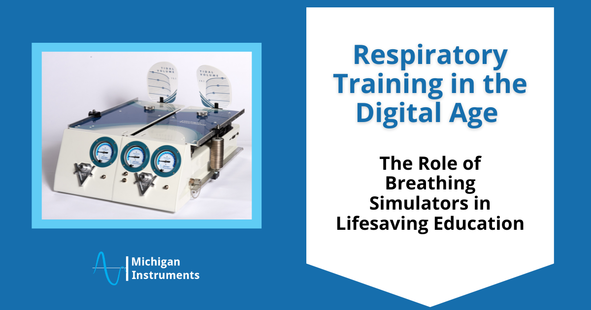
Educators, technicians, and researchers need to equip their departments with the right tools to effectively train and test respiratory care techniques. Lung Simulators offer some of the most advanced training and testing capabilities available today.  This effectively helps respiratory students and professionals bridge the gap between theoretical knowledge and real-world application.

Advanced Training Test Lungs for Comprehensive Learning

But not all Lung Simulators are created equal! Unlike more basic test lungs, Michigan Instruments’ Training Test Lungs offer residual lung volumes and a dynamic response to therapy that more closely mirrors actual patient conditions. 

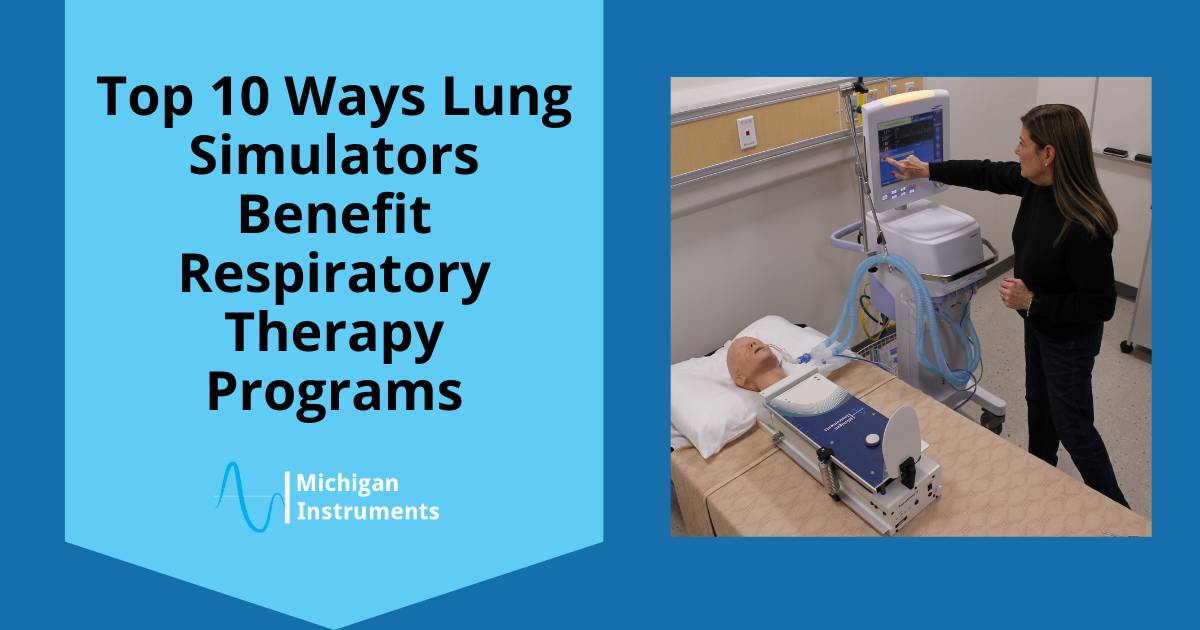
Michigan Instruments Training Test Lungs are designed to realistically simulate a wide range of healthy and diseased lung conditions by offering adjustable settings for lung compliance and airway resistance. These features provide an accurate representation of the human pulmonary system, and allow the students invaluable practice with mechanical ventilators, BVMs, and other respiratory equipment before they even enter the clinical setting. 

Effective Respiratory Training and Real-World Applications

Many of the Respiratory Therapy training programs across the U.S. and  around the world use Michigan Instruments’ TTL and PneuView® systems because they are recognized for their versatility, ease of use, and durability, making them essential tools in education and research alike.

Michigan Instruments Training Test Lungs are a better choice because they:

  • Help students understand complex respiratory issues by simulating a wide range of lung conditions.
  • Allow students to work with a realistic total lung and residual volumes.
  • Incorporate many options and great versatility regarding tapping into the “lungs” to introduce gasses, perform additional monitoring or connect to auxiliary devices
  • Simulate asymmetrical lung disease and the resulting ventilation dynamics with The Dual Adult TTL and Dual Adult PneuView systems.
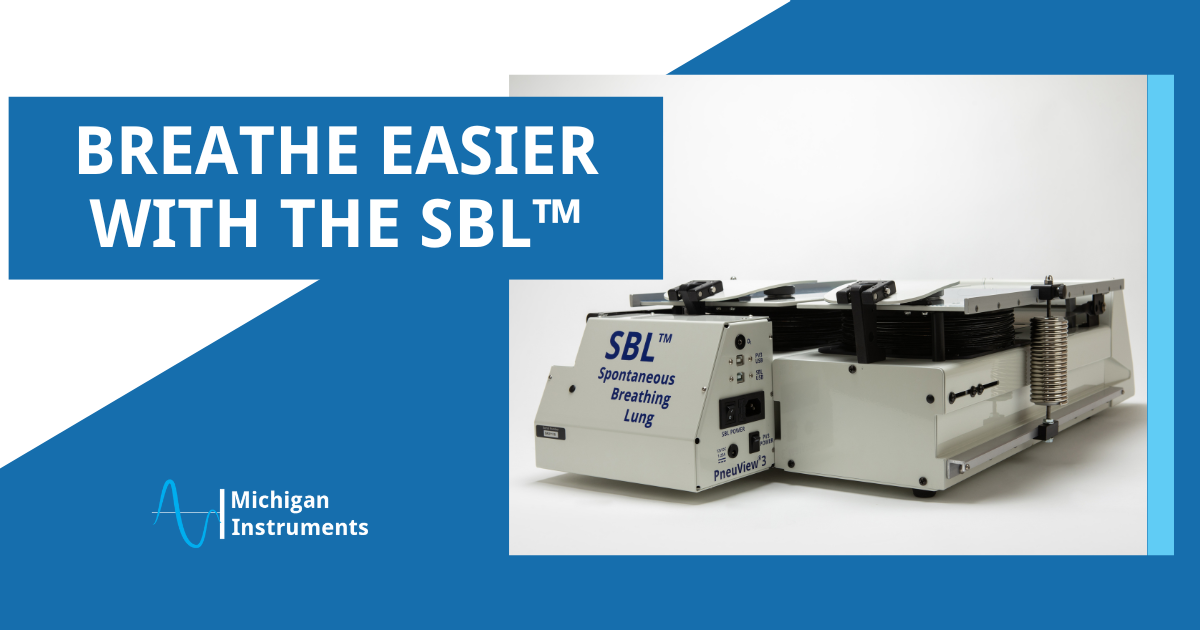
  • Simulate a spontaneous breathing patient and evaluate the response of devices in various support modes.
  • Facilitate education and studies with various oxygen delivery systems, non-invasive ventilation devices/techniques, and other applications requiring a “face” with available Head Simulation Modules 
  • Are designed to be versatile and durable to easily withstand repeated student use in demanding classroom settings.

Train with PneuView® Software

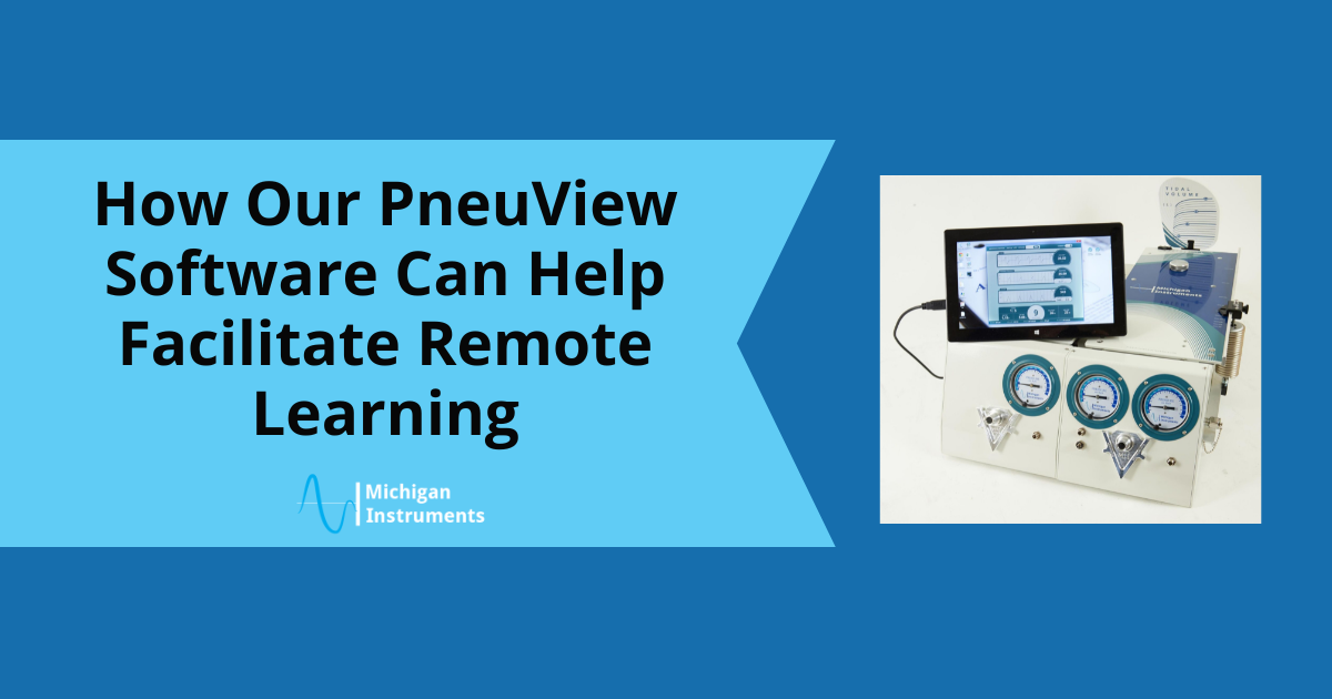
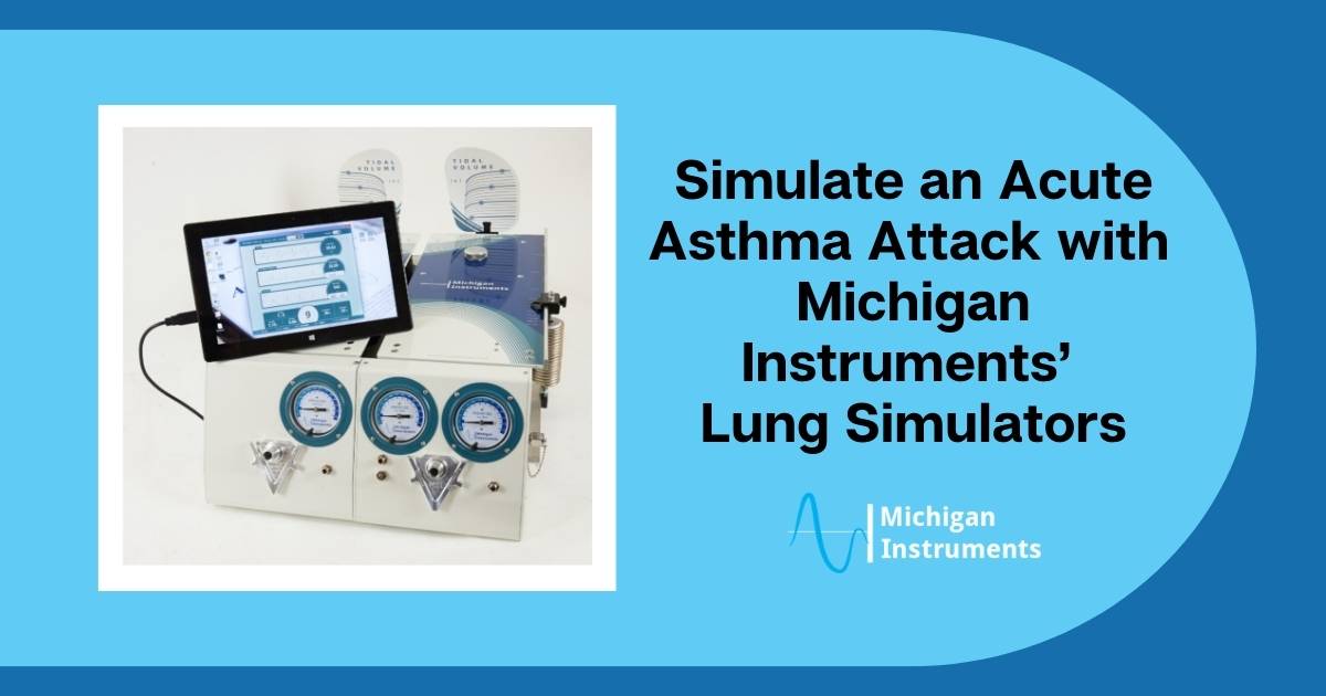
With the PneuView® system, educators and students can take their training even further! This PC-based software interacts with our lung simulators to display real-time data, including waveforms for pressure, volume, and flow. Even when using simple ventilation devices like BVMs, CPAP systems, or emergency ventilators, PneuView allows for dynamic feedback, helping students analyze performance and patient responses.

PneuView can also be used to view, capture, record, and replay ventilation data, making it easy to save and export data for later review. This feature is perfect for storing recordings of ventilation runs, allowing instructors to demonstrate and evaluate techniques during post-session reviews. It’s also invaluable for students and professionals conducting research projects, as the software simplifies data collection and analysis.

How Do These Devices Benefit Your Students?

The TTL® and PneuView® Lung Simulators provide an unmatched combination of hands-on experience, real-time feedback, and adjustability. Students can gain invaluable experience by practicing with simulated pulmonary mechanics that mimic the real-world response to various devices and therapies. By using these tools, training programs can elevate the quality of education they provide, ensuring that students are better prepared to handle real-life respiratory care challenges.

Real-World Testimonials from Educators

Michigan Instruments’ Training Test Lungs and PneuView®3 systems have become invaluable tools for educators and professionals in the respiratory care field. Here’s what some of our users have to say.

Tim Op’t Holt highlights the versatility of the TTL for both education and research, showcasing how the simulators provide realistic feedback for students and drive important studies in respiratory care.

“We use the Michigan Instruments Test Lungs / Lung Simulators (TTL) in our program for demonstration of mechanical ventilation, laboratory practice with ventilators, and aerosol and ventilator research. The TTL is valuable in showing students the effects of changes in compliance and resistance on volume and pressure… In research, we use the breath simulation module to drive the left lung which lifts the right lung to act as a breathing lung for aerosol deposition studies.”  

Tim Op’t Holt EdD, RRT, AE-C, FAARC
Professor and Chair, Cardiorespiratory Care – University of South Alabama


Debbie Grube emphasizes the accessibility and affordability of Michigan Instruments’ Test Lungs, underscoring their importance in a classroom setting where advanced simulation technology may not always be readily available.

“Our Respiratory Therapy Program currently owns 4 of the Michigan Instruments TTL® Lung Simulators… The Michigan Instruments lungs are tremendously useful tools that can be easily manipulated by our students, are cost-effective, and are readily available in our own classroom. We consider the TTL’s an essential part of our training program!”  

Debbie Grube RRT, MSM
Director of Respiratory Care – Muskegon Community College


Dr. Ann Flint points out the durability and ease of use of Michigan Instruments’ devices, making them essential tools for a wide range of lab exercises and real-world training.

“Michigan Instruments has the best test lung to easily provide simulation of a variety of lung conditions that even students can adjust… They are versatile, and a great tool for giving our students hands-on experience with mechanical ventilation techniques, simulation of disease states, performing experiments to test various concepts, and much more. They are easy to use and durable. We wish we had more!”  

Dr. Ann Flint
(Former) Program Director, Respiratory Care – Jackson College

Breathe New Life into Your Classroom With Michigan Instruments

At Michigan Instruments, our goal is to support respiratory education and research by providing advanced simulation tools that help bridge the gap between theory and practice. Our TTL® and PneuView® Lung Simulators are designed to enhance your training program, offering realistic lung simulations, versatile applications, and easy-to-use software that prepares students and professionals alike for real-world scenarios.

If you’d like to learn more about how these devices can benefit your program, contact us today.

personalized healthcare

Personalization has become a growing trend in many industries, with consumers seeking tailored experiences in everything from their skincare routines to their water bottles. While this trend has taken off in the consumer world, its impact is even more profound in healthcare. 

The Personalized Medicine Revolution

Personalized medicine, which tailors decisions and treatments related to an individual’s unique needs using their genetic profile, is transforming the medical landscape. Personalized medicine changes healthcare from a “one size fits all” approach to an individualized approach, making decisions for each person based on their specific molecular makeup. 

This evolution of healthcare has been made possible by enabling technologies, such as nanotechnology for isolating extracellular vesicles and imaging platforms to predict nanomedicine’s effectiveness. Technologies like these have shifted the landscape of healthcare, offering diagnostic insight on a personalized level like never before.

The Power of Lung Simulators in Personalized Medicine

Respiratory care has also been impacted by the evolution of personalized healthcare, and tools like lung simulators are leading the charge. In the respiratory space, healthcare professionals are able to customize treatments for each patient’s specific condition, size, and age  This remarkable potential extends even beyond human applications. 

Lung simulators replicate the mechanics of human lungs and can be adjusted according to a number of factors and different combinations, such as lung capacity, compliance, and airway resistance.

By mimicking a variety of lung conditions, lung simulators provide healthcare professionals with an inside look into how different treatments interact with various patient conditions. With this information at their fingertips, physicians can develop more precise treatment plans based on real-time data, reducing the risks associated with trial-and-error approaches. 

Customizing Lung Simulators for Diverse Patient Needs

Michigan Instruments’ lung simulators allow clinicians to adjust settings such as tidal volume, respiratory rate, and airway pressures to reflect a range of patient profiles, ensuring that treatments are truly personalized to an individuals’ specific profile for optimal results.

Tailoring for Age and Size

Infants, children, adults, and elderly patients all have distinct respiratory needs. A Michigan Instruments lung simulator can be set to replicate the lung dynamics for a wide range of age related conditions, helping healthcare providers customize ventilator settings for the specific needs of pediatric or geriatric patients.  

Condition-Specific Simulation

Lung diseases like COPD, pulmonary fibrosis, or even acute conditions like pneumonia each present their own unique challenges. Lung simulators allow healthcare professionals to simulate these conditions, which helps them fine-tune treatments before applying them to patients. 

This capability is particularly crucial for managing patients with chronic conditions that require long-term care and frequent adjustment of ventilator settings.

Beyond Human Applications

While lung simulators are primarily designed to enhance human healthcare, their versatility extends beyond human applications. In a unique and extraordinary case, Michigan Instruments was called upon to create a specialized lung simulator for a dolphin. 

By successfully creating a dolphin-specific lung simulator, we demonstrated the flexibility and precision of the lung simulator technology, proving that simulators can be customized to suit even the most unconventional respiratory systems and highlighting the broader applications of lung simulation technology.

Lung Simulators in Research and Training

The use of lung simulators goes beyond direct patient care, playing a significant role in medical research and training. By providing a controlled environment where medical professionals can study how different therapies interact with diverse patient conditions, these simulators help to advance the field of respiratory medicine and personalized medicine. 

Medical teams can simulate challenging scenarios, such as dealing with patients who have complex comorbidities, allowing them to develop and practice personalized treatment strategies. This level of preparation ensures that patient care is both safer and more effective, as clinicians can test and refine their approaches before they ever reach the patient’s bedside.

The Impact on Critical Care and Emergency Medicine

While most personalized healthcare is focused on chronic or long-term conditions and treatments, it can also be applied to emergency and critical care settings. For patients experiencing acute respiratory distress or failure, time is of the essence. Lung simulators combined with personalized medicine can help healthcare providers optimize ventilator settings quickly and accurately, based on patient-specific conditions.

In trauma care or emergency medicine, lung simulators provide a deeper understanding of how different ventilation strategies can affect patient outcomes. This level of precision is key in critical care scenarios where every second counts, and the ability to personalize care can make the difference between life and death.

The Role of CPR Devices: Enhancing Personalized Care

While lung simulators represent the cutting edge of respiratory care, other life-saving technologies complement the effort to deliver personalized medicine. For instance, automated CPR is another opportunity for customization based on individual patient needs.

Automated CPR devices are designed to provide consistent, high-quality chest compressions and ventilations during resuscitation efforts, but they can also be adapted based on the patient’s size, weight, and condition. 

For instance, a smaller adult can require a different compression depth than a larger individual, and these devices can be adjusted accordingly – increasing or decreasing the depth of compression. In addition, the ventilation settings on devices like the Life-Stat can be  set to offer 2 breaths automatically after 30 compressions or 9 asynchronous breaths during continuous compressions.

Though they are primarily used in emergency situations, these CPR devices reflect the same commitment to personalized care that lung simulators provide. Together, they ensure that patients receive treatments tailored to their specific needs, whether in the emergency/hospital rooms or during long-term respiratory therapy.

Advancing Personalized Medicine with Lung Simulators

As healthcare continues to evolve toward a more individualized approach, Michigan Instruments’ lung simulators stand at the forefront of this movement. By enabling clinicians to simulate a wide variety of patient conditions and tailor treatments accordingly, these devices are helping to improve patient outcomes and push the boundaries of what personalized medicine can achieve.


We provide a comprehensive suite of tools designed to deliver the most effective, patient-specific care possible. The future of respiratory care is personalized, and lung simulators are helping to lead the way.

Interested to learn more? Contact us today.

ventilator associated lung injury

In most cases, ventilators save lives—but like any other medical procedure, it doesn’t come without risks. Ventilator-associated lung injuries (VALI) occur when improper ventilation techniques are used, and can result in pulmonary edema, barotrauma, or worsening hypoxemia that can prolong mechanical ventilation. 

In the worst cases, VALI can also lead to multi-system organ dysfunction and increase the chances of mortality in the patient. 

Best Practices to Minimize the Risk of Ventilator-Associated Lung Injuries 

To minimize the risks of complications, it’s essential to follow strict protocols and best practices while placing and managing ventilated patients. 

Below, we lay out key strategies to prevent VALI and highlight the role of advanced training tools in ensuring safe and effective ventilation.

What Are Ventilator-Associated Lung Injuries?

Ventilator associated lung injury occurs when mechanical ventilation, though life-saving in most cases, inadvertently causes harm to the lung tissue. This damage can lead to complications with serious consequences. Common types of VALI include:

  1. Pulmonary edema: When fluid collects in the many air sacs in the lungs making it difficult to breathe. Pulmonary edema may lead to death.
  2. Pulmonary barotrauma: When the air pressure in the lungs changes too quickly, causing damage to the lung tissue. Pulmonary barotrauma can lead to a collapsed lung or death. 
  3. Worsening hypoxemia: When oxygen levels in the blood are lower than normal, which can interfere with heart and brain function. It can also lead to a lack of oxygen in your body’s organs and tissues.
  4. Multi-system organ dysfunction: When two or more organ systems are failing. It’s a serious medical emergency that’s fatal without immediate treatment.  

Each of these injuries can make the patient’s condition much worse, leading to a prolonged hospital stay, requiring further life-saving measures, and even increasing the risk of mortality. It’s clear that proper management of ventilator settings, comprehensive monitoring, and an awareness of patient-specific factors is crucial.

What are the Best Practices in Ventilator Management?

Most hospitals and/or medical providers have best practices in place for every procedure and process performed. Ventilator management is one of the most important processes in life-saving situations, so adopting best practices related to ventilation is crucial. 

Luckily, there are a number of well-established best practices in mechanical ventilation management that can help keep patients safe during ventilation:

  1. Low Tidal Volume Ventilation (LTVV): Using lower tidal volumes (typically 4-8  mL/kg of ideal body weight) is crucial to reducing the risk of lung overdistention and other complications. 
  2. Optimal Positive End-Expiratory Pressure (PEEP): At the end of mechanical or spontaneous exhalation, PEEP maintains the patient’s airway pressure above the atmospheric level by exerting pressure that opposes passive emptying of the lung. Ensuring safe yet effective PEEP levels can also decrease the risk of VALI. 
  3. Limiting Plateau Pressure: Maintaining plateau pressures below 30 cm H₂O helps prevent barotrauma, which is caused by excessive pressure on the lungs. Regular monitoring ensures ventilation settings are optimal for the patient’s condition.​
  4. Individualized Ventilation Strategies: Tailoring ventilation settings to the specific lung mechanics and disease state of each patient helps ensure both effective and safe ventilatory support​.
  5. Early Mobilization and Sedation Management: Reducing sedation as early as safely possible and promoting early mobilization can help shorten the duration of mechanical ventilation, reducing the risk of associated complications.

Strategies to Prevent Ventilator-Associated Lung Injuries 

In addition to the above established protocols and best-practices, healthcare providers can adopt other strategies to enhance the safety and efficacy of mechanical ventilation, such as:

  • Continuous Monitoring: Utilize advanced monitoring tools to assess lung mechanics, gas exchange, and patient-ventilator interaction in real-time to make the best decisions for each individual patient.
  • Interdisciplinary Collaboration: Ensure that respiratory therapists, nurses, and physicians work closely together to optimize ventilatory support and adjust settings as needed.
  • Regular Training and Simulation: Ongoing education and hands-on training in ventilator management are essential to keep healthcare professionals up-to-date with the latest techniques and technologies.

How to Enhance Confidence and Precision in Providers and Reduce Ventilator-Associated Lung Injuries

While all of the previously mentioned strategies are critical in reducing ventilator associated lung injury, it’s crucial for medical organizations to recognize and remember that proper, ongoing training is a great way to prevent ventilator-associated lung injuries. 

Why Training Matters

Hands-on experience with lung simulators and ventilation products enables doctors and respiratory therapists to gain a deeper understanding of lung mechanics and the complexities of ventilator management. 

Personnel, technologies, and techniques are regularly changing and evolving. Lung Simulators allow caregivers to get comfortable with new techniques and devices in a risk-free environment prior to applying them to actual patients. 

How to Build Confidence

Regular training with the addition of lung simulators helps healthcare professionals become more confident in placing ventilators and adjusting settings to suit individual patient needs. This confidence translates to better patient outcomes, as clinicians are more adept at avoiding common pitfalls associated with mechanical ventilation.

How to Practice Precision in Care

Accurate placement and management of ventilators are critical in preventing VALI. By using training models and products, healthcare professionals are provided the opportunity to practice the precision needed to ensure that ventilatory support is both safe and effective, reducing the likelihood of lung injuries and improving overall patient care.

Prevent Ventilator-Associated Lung Injury with Michigan Instruments 

Preventing ventilator-associated lung injuries requires a combination of following established protocols, engaging in ongoing education, and the use of advanced training tools. 

By investing in comprehensive training with Michigan Instruments’ products, such as the Training Test Lung, healthcare professionals can enhance their skills, boost their confidence, and deliver the highest standard of care to their patients.

Contact us to learn more.

modern medical education

The landscape of education is evolving rapidly, and modern medical education is no different! Medical schools around the world are embracing innovative strategies and technologies to enhance students’ learning experiences and prepare the next generation of healthcare professionals for all that they will encounter. 

In this blog, we’ll explain some of these cutting-edge technologies and strategies.

The Best New Ways to Learn in Medical Education

The newest teaching methods focus on hands-on training, interactive learning, and real-world simulations, ensuring that medical students and professionals are well-equipped to handle all of the complexities of modern healthcare. 

Interdisciplinary Team Training

Interdisciplinary team training emphasizes the importance of collaboration and communication among healthcare professionals from different specialties. While this hasn’t been common practice in the past, this approach mirrors the real-world dynamics of healthcare settings, where teamwork is essential for delivering high-quality patient care.

Realistic Lung Simulation

Realistic lung simulators are particularly well suited for medical education and training.  These simulators provide realistic respiratory simulation. As the Lung Simulators mimic the human pulmonary mechanics, the students can simulate a wide array of healthy and diseased lung physiologic conditions. This allows the students the hands-on experience to interpret data, manage technologies and learn new therapies.

Flipped Classroom Model

The flipped classroom model reverses the traditional learning environment by delivering instructional content online, outside of the classroom. This allows class time for interactive, hands-on activities and problem-solving.

This approach allows students to learn at their own pace and use classroom time more effectively for engaging discussions, collaborative projects, and practical application of knowledge.

Virtual Reality and Augmented Reality

Virtual Reality (VR) and Augmented Reality (AR) technologies are transforming modern medical education by providing immersive and interactive learning experiences. These technologies enable students to visualize complex anatomical structures, practice surgical procedures, and engage in patient interaction– all virtually.

Problem-Based Learning

Problem-based learning (PBL) encourages critical thinking and problem-solving skills by presenting students with real-world medical cases. This student-centered approach aids in independent learning and application of knowledge to practical scenarios.

Telemedicine Training

Telemedicine training prepares medical students to provide care remotely using telecommunication technology. This training includes virtual consultations, remote monitoring, and the use of digital health tools.

Telemedicine training equips future healthcare professionals with the skills needed to deliver care in a rapidly evolving healthcare landscape. It also expands access to care, especially in rural or underserved areas.

3D Printing and Anatomical Models

3D printing technology is used to create detailed anatomical models that students can use for hands-on learning. These models can replicate complex structures, such as organs and tissues, providing a tactile learning experience.

3D printed models allow students to explore anatomy in a way that is not possible with traditional textbooks. They provide a deeper understanding of spatial relationships and structures within the human body.

Remote and Online Learning

It’s no secret that the COVID-19 pandemic accelerated the adoption and necessity of remote and online learning in medical education. However, even now, virtual classrooms, online simulations, and telemedicine training are integral components of medical curricula, ensuring continuity of education regardless of physical location.

How Michigan Instruments is Preparing the Next Generation of Healthcare Professionals

Michigan Instruments’ Training Test Lungs are essential tools for remote learning, PBL sessions, and simulation labs—offering students hands-on experience with ventilation techniques and respiratory mechanics. 

Students can use the test lungs to explore various clinical scenarios, such as managing different lung compliance and resistance conditions. This hands-on approach helps learners understand the principles of mechanical ventilation and develop problem-solving strategies for respiratory care. 

When integrated with VR simulations, students can practice intubation, ventilation, and other respiratory procedures in a virtual environment. Observing and interacting with these devices in real-time provides students with immediate feedback, making respiratory training both effective and engaging. 

Our Automated CPR Devices are also excellent tools for team training exercises. During cardiac arrest simulations, students from various disciplines—such as emergency medicine, nursing, and respiratory therapy—can work together to practice coordinated responses. The automated CPR device ensures consistent chest compressions, allowing the team to focus on other critical tasks and improve their collaborative skills.

Join Us in Changing the Landscape of Medical Education

The integration of cutting-edge teaching strategies in medical education is essential for preparing healthcare professionals to meet the demands of modern medicine. 

Our innovative products play a pivotal role in enhancing these educational experiences, providing realistic, hands-on training that bridges the gap between theory and practice. As medical education continues to evolve, the collaboration between educational institutions and medical device manufacturers like us will be crucial in shaping the future of healthcare.

Contact us today to see how our products can elevate your medical training programs and prepare the next generation of healthcare professionals.