history of respiratory therapy

The computer-controlled ventilators we know and use today operate on basic principles that are more than a century old. While the history of respiratory therapy dates back, there’s no denying that the advancements in the field have come a long way since then! 

What is Respiratory Failure?

To fully understand the miracle of respiratory therapy, we first need to grasp the gravity of respiratory failure. 

Respiratory failure is a condition that makes it difficult for a person to breathe on their own, resulting in the lungs’ inability to supply blood with oxygen. As breathing is an essential function of life; without sufficient breathing, our tissues and organs cannot work properly. 

According to NHIBI, when we inhale oxygen from the air into our lungs, we exhale out carbon dioxide, which is a waste gas made in the body’s cells. Improper breathing leads to a buildup of carbon dioxide. This buildup can damage tissues and organs and slow oxygen delivery to the body.

Some of the different causes of respiratory failure include:

  • Nerve and muscle disorders
  • Lung and airway diseases 
  • Fluid buildup in the lungs or pulmonary embolism 
  • Infections in your brain or spinal cord, lungs, or airways
  • Blocked airway when food or another object gets stuck in your airways
  • Chest or back injuries
  • Severe scoliosis
  • Severe allergies to food or medicine
  • Lifestyle habits, like smoking or using drugs or alcohol 

The Ventilator: The Medical Device You Hope You’ll Never Need

When encountering respiratory failure, the use of a ventilator is an extreme life-saving measure.  These are used when a patient’s lungs are not functioning properly on their own. Ventilators give a patient’s body a much-needed rest in addition to support, allowing their bodies to heal.

The use of ventilators goes back quite some time, and there are different types of ventilators on the market today. But how has technology changed over the years? 

Whole-Body Ventilators Lead to The Iron Lung

In an article written by Robert M Kacmarek, it is noted that the negative-pressure ventilator was the predominant device used to provide ventilatory assistance in the 19th and early 20th centuries. 

  • 1838: The earliest ventilator was described as a full body ventilator, or as a “tank ventilator”. The ventilator was an air-tight box, with the patient in a sitting position while negative pressure was established by manually pumping air into and out of the box. 
  • 1904: Following the Tank Ventilator were similar types of manually operated negative-pressure ventilators, like Sauerbrach’s negative-pressure operating chamber in 1904. In this chamber, the patient’s body, except for the head, was maintained inside a chamber which was large enough so that the surgeon was able to perform surgery while also in the chamber. 
  • 1907: Johann Heinrich Dräger and his son Bernhard introduced The Pulmotor, one of the only early devices for positive pressure ventilation in 1907. The Pulmotor was a transportable device that used a face mask to deliver oxygen until a set pressure was reached in the lungs, at which point the device switched to assisting the patient with exhalation. 
  • 1928: The Iron Lung, originally designed and built by Drinker and Shaw, but manufactured and sold by Emerson, was one of the most widely used ventilation devices of the early 20th century. It used negative-pressure techniques and worked by changing the pressure inside an airtight container. It expanded and contracted the chest, pulling air in and out of the lungs, and providing much needed support in the ICU for children suffering from polio for decades.

Advancements in Positive Pressure Ventilation are Made 

Just after the Polio epidemic of the 1950s, there was a movement away from negative-pressure ventilation.

This was mostly due to:

  1. Volume-targeted ICU/anesthesia ventilators beginning to appear. 
  2. The development of small, compact, intermittent positive-pressure breathing (IPPB) devices.
  3. The use of negative-pressure ventilation became too much, as they were large, heavy, and cumbersome.

The development of new positive-pressure breathing devices is credited to the need for pilots of late-WWII era jet planes requiring oxygen for their flights at high altitudes.

In the mid-1950’s, the Bird Mark 7 Respirator was developed by a former U.S. Army pilot, Forrest Bird. Many consider this device to be the first modern medical respirator, which led to many advancements in ventilator design and technology.

Non-Invasive Ventilators Through the Years

Though negative pressure, whole-body ventilators were most widely used at first, other non-invasive ventilators existed as well.

  • 1780: The bag and mask manual ventilator is introduced by Chaussier
  • 1887: A more sophisticated bellows with a mask was introduced by Fell
  • 1907: Dräger’s Pulmotor was first introduced, and for its time, was fairly sophisticated. The pneumatically operated positive-pressure device has been credited with saving thousands of individuals over its lifetime.
  • 1910: Green and Janeway create a device known as a “rhythmic inflation apparatus.” The patient’s head was placed into the apparatus and a seal was secured around the patient’s neck with positive pressure applied to the patient’s head. 
  • 1957: The Bird Mark series of ventilators were originally created to provide intermittent breathing treatments as opposed to long-term ventilation. Soon after, they began to be used for life support in both noninvasive and invasive ventilation.

Continued Advancements in Positive Pressure Ventilation Leads to Invasive Options

Another key part of the history of respiratory therapy is positive pressure ventilation. The development of ventilators for positive-pressure invasive ventilation began in the 1940s, and with each generation, became more advanced. Early models provided volume-control ventilation but lacked patient-triggered ventilation. Examples included the Morch ventilator, a simple piston ventilator, and the Engstrom ventilator, more sophisticated with double-circuit capabilities. 

The first-generation ventilators didn’t incorporate PEEP until the 1970s. The second generation introduced patient-triggered inspiration and basic alarms like high pressure and low tidal volume. Intermittent mandatory ventilation (IMV) emerged during this period. 

Third-generation ventilators, with microprocessor control, marked a significant advancement with extensive monitoring, waveforms, and various ventilation modes like pressure support and SIMV. 

Fourth-generation ventilators are the most complex, featuring numerous ventilation modes, closed-loop control, and capabilities for non-invasive ventilation (NIV) and neonatal ventilation. They also offer extensive monitoring and specialized management packages for weaning and assessments.

Michigan Instruments is Committed to Continued Advancements

Michigan Instruments Lung Simulators play a crucial role in advancing ventilator technology through various contributions that enhance training, testing, research, and product development in the field of respiratory therapy.

  • Training: We offer the Spontaneous Breathing Lung (SBL™), a sophisticated device that facilitates training for healthcare professionals and students. With the SBL™, users can create a wide range of breathing scenarios and patterns, making it an invaluable tool for educating professionals on new respiratory devices, ventilators, and therapeutic modalities.
  • Testing: The SBL™ is instrumental in testing and troubleshooting devices designed for spontaneous breathing patients. It enables thorough assessments, such as detecting the start of a breath and avoiding breath-stacking, ensuring the optimal performance of respiratory devices.
  • Research & Development: Our Lung Simulators are highly versatile for research applications. They have been utilized in diverse studies, from analyzing aerosol dispersion to evaluating the responsiveness of new ventilation. Researchers benefit from the precise simulation capabilities of Michigan Instruments’ equipment, enhancing the quality and accuracy of their research outcomes.

The SBL™ offers advanced control over breath rate, tidal volume, inspiratory time, and inspiratory flow pattern. Additionally, it has customizable settings and intuitive software, so users can simulate spontaneous breathing accurately. This makes it ideal for designing, testing, and training on non-invasive ventilation and oxygenation modalities.

Elevate Your Research Abilities with Michigan Instruments

Our commitment to innovation and excellence in respiratory care is evident in our comprehensive range of simulation tools and software solutions. We continue to empower healthcare professionals, educators, and researchers worldwide—contributing significantly to the evolution of ventilator technology and respiratory therapy practices.

For more information on our devices used in research environments worldwide, contact us today.

how to prevent lung disease

The lungs are undoubtedly one of the most important organs in the human body. They play a vital role in our overall health and well-being while serving as the gateway for oxygen intake and the removal of carbon dioxide. Their function is essential to sustaining life itself. 

Beyond that, the lungs actively participate in immune defense, support physical activity, and contribute to the overall balance of the body’s systems. Understanding the significance of lung health and function goes beyond breathing; it supports our capacity to thrive and maintain optimal health. 

Keep reading to know more about the importance of lung health and how to prevent lung disease.

The Importance of Lung Health and Function

In this blog, we’ll take a closer look at the importance of lung health and function. We’ll explore how these critical organs impact every aspect of our lives, and why nurturing them is essential for our longevity and quality of life.

Maintaining optimal lung health is crucial for several reasons:

1. Oxygenation of the Body

The primary function of the lungs is to supply oxygen to the bloodstream and remove carbon dioxide. Adequate oxygenation is essential to ensure proper function of all your organs. 

Without oxygenation, cells can’t perform their functions, which leads to inadequate overall health.

2. Respiration

Breathing is an involuntary action controlled by the respiratory system. Your lungs expand and contract with each breath, allowing air to flow in and out of the body. 

Efficient respiration ensures that oxygen is delivered to tissues and organs and waste gasses are eliminated, supporting metabolic processes and maintaining a healthy pH balance.

3. Immune Defense

Did you know that your respiratory system plays a crucial role in defending the body against pathogens, pollutants, and foreign particles? 

It’s true; mucus and cilia in the airways trap and remove harmful substances. Immune cells in the lungs detect and neutralize invading microorganisms, helping to prevent infections and maintain your respiratory health.

4. Physical Activity and Endurance

Healthy lungs allow you to engage in physical activities and exercise without experiencing fatigue or shortness of breath. Strong respiratory muscles and efficient breathing gives you endurance and stamina to lead an active and fulfilling life.

5. Overall Health and Well-being

Lung health is closely linked to overall health and well-being. Chronic lung conditions such as COPD, asthma, and lung cancer can significantly impact your quality of life.  These conditions can lead to symptoms such as coughing, wheezing, and difficulty breathing. Maintaining optimal lung function is essential for preserving physical and mental health and enjoying a high quality of life.

It’s clear that lungs are vital for sustaining life, supporting physical activity, and protecting against respiratory illnesses and diseases. By taking proactive steps to maintain respiratory health, you can enhance your overall well-being and enjoy a higher quality of life.

Understanding Lung Diseases

Learning how to prevent lung disease starts with understanding some of the most common ones.  Human lungs are susceptible to various diseases that can impact our quality of life. Let’s explore the 7 most common lung conditions and discuss strategies to prevent them.

Chronic Obstructive Pulmonary Disease (COPD)

COPD is a progressive lung disease characterized by obstructed airflow, making it difficult to breathe. It encompasses conditions such as chronic bronchitis and emphysema, often caused by long-term exposure to irritants like cigarette smoke or air pollution. 

Symptoms include coughing, wheezing, and shortness of breath, which can significantly impact daily activities and quality of life.

Asthma

Asthma is a chronic respiratory condition characterized by inflammation and narrowing of the airways, leading to recurrent episodes of wheezing, coughing, chest tightness, and shortness of breath. 

Daily triggers such as allergens, respiratory infections, and environmental factors can make symptoms worse, making it essential for individuals with asthma to manage their condition effectively.

Lung Cancer

Lung cancer is one of the most common yet deadliest forms of cancer worldwide.  It  is often linked to smoking, subjection to secondhand smoke, and exposure to carcinogens like asbestos and radon. 

Early cancer detection through screenings plus adopting a healthier lifestyle can improve outcomes and increase survival rates for those diagnosed with lung cancer.

Pneumonia

Pneumonia is an infection that inflames the air sacs in one or both lungs, causing them to fill with fluid or pus. It can be caused by bacteria, viruses, or fungi and is characterized by symptoms such as fever, chills, cough, and difficulty breathing. 

Pneumonia can range from mild to severe and may require hospitalization, particularly in vulnerable populations such as the elderly and individuals with weakened immune systems.

Pulmonary Fibrosis

Pulmonary fibrosis is a progressive lung disease characterized by scarring of the lung tissue, which makes it difficult for the lungs to function properly. While the exact cause is often unknown, factors such as exposure to environmental toxins, certain medications, and autoimmune diseases may contribute to its development. 

Symptoms include shortness of breath, dry cough, fatigue, and unexplained weight loss, with treatment aimed at managing symptoms and slowing disease progression.

Bronchiectasis

Bronchiectasis is a chronic condition characterized by permanent enlargement of the airways in the lungs, leading to recurrent infections and inflammation. It can occur alongside conditions such as cystic fibrosis, respiratory infections, or inhaling toxic substances. 

Symptoms include persistent cough, excess mucus production, and frequent respiratory infections, with treatment focusing on clearing airway secretions, managing infections, and preventing complications.

Pulmonary Embolism

A pulmonary embolism occurs when a blood clot travels to the lungs and blocks blood flow, leading to potentially life-threatening consequences. Risk factors include immobility, surgery, pregnancy, and certain medical conditions such as cancer and thrombophilia. 

Symptoms can vary but may include sudden onset of chest pain, shortness of breath, rapid heart rate, and coughing up blood. Prompt medical attention is essential to prevent complications and improve outcomes for individuals with pulmonary embolism.

Preventing Lung Diseases

The lung diseases outlined above can lead to decreased quality of life and even death. It’s important to care for our lungs to lead a healthy, fulfilling life. 

While some lung diseases are unavoidable due to environmental factors or because of other diseases, preventing most lung diseases is possible by leading a healthy life.

Here are some ways to prevent these lung diseases.

1. Avoid Tobacco Smoke

Tobacco smoke is a leading cause of lung disease and cancer. If you smoke, quitting is the single most important step you can take to protect your lung health. 

Avoid exposure to secondhand smoke and encourage others to do the same, especially around children and individuals with respiratory conditions.

2. Maintain a Healthy Lifestyle

Eating a balanced diet rich in fruits, vegetables, and whole grains can support lung health by providing essential nutrients and antioxidants. Regular exercise strengthens the respiratory muscles and improves lung function, reducing the risk of lung disease and promoting overall well-being.

3. Minimize Exposure to Air Pollutants

Indoor and outdoor air pollutants, such as particulate matter, ozone, and nitrogen dioxide, can contribute to respiratory problems and worsen existing lung conditions. Take steps to minimize exposure by using air purifiers, avoiding outdoor activities during high pollution days, and advocating for clean air policies in your community.

4. Practice Good Hygiene

Practicing good hygiene , such as washing hands frequently, covering coughs and sneezes, and staying home when sick, can help prevent the spread of respiratory infections like influenza and COVID-19. 

Additionally, getting vaccinated against preventable respiratory illnesses can provide added protection for yourself and those around you.

Safeguarding Our Lungs is Key to Optimal Health

The importance of good lung health and function cannot be overstated. From the oxygenation of our bodies to immune defense and physical endurance, our lungs are indispensable to our well-being. 

By understanding the common conditions that can affect lung health and adopting preventive measures, we can safeguard these vital organs and enhance our overall quality of life. 

Whether it’s avoiding tobacco smoke, maintaining a healthy lifestyle, or practicing good respiratory hygiene, every action we take contributes to the health and longevity of our lungs. 

Join Michigan Instruments in prioritizing lung health, not only for ourselves but for future generations, ensuring that we can continue to breathe easy and thrive in the years to come.

Exploring Lung Health Innovations with Michigan Instruments

At Michigan Instruments, we’re committed to advancing respiratory care. Our TTL and PneuView Lung Simulators serve as indispensable tools in the realm of medical research and development. These simulators offer realistic and precise simulation capabilities, providing engineers with a safe and controlled environment to design and test therapeutic and monitoring devices.

Our commitment to respiratory research and care isn’t solely for human use: In a 2018 collaboration with a veterinary facility, we embarked on a unique project to simulate dolphins under anesthesia. 

By developing and testing a specialized solution tailored to their needs, we helped ensure the safety and efficacy of their dolphin ventilator for medical use.

The Future of Lung Health Begins Here

With options for setup, accuracy, reliability, and durability, our lung simulators empower researchers to achieve consistent settings and repeatable results. The integration of PneuView software further enhances the capabilities of our simulators, allowing R&D professionals to access real-time data collection for documentation, review, and analysis.

Our fully calibrated lung simulators are available in single or dual-lung models for both adults and infants, offering researchers the flexibility to explore a wide range of healthy and diseased lung conditions. 

Additionally, our Spontaneous Breathing Lungs enable the simulation of natural breathing patterns, enhancing the realism of research scenarios.

Request a quote for our different devices including the lung simulation products or mechanical CPR devices (Thumper & Life-Stat).

respiratory simulation

Technological advancements continue to shape the way professionals acquire critical skills in medical education and research. 

One such innovation that stands out is the integration of respiratory training simulators, specifically lung simulators, into the educational toolkit of medical professionals, ventilator manufacturers, and educators. 

Among the pioneers in this field is Michigan Instruments, offering state-of-the-art lung simulators paired with PneuView Software.

What Sets Our Lung Simulators and PneuView Software Apart?

Michigan Instruments takes pride in providing lung simulators that are fully to scale, offering realistic residual lung volumes and capacities. The range of settings for compliance and resistance on TTL® and PneuView® systems surpasses most other available simulators. What makes these devices truly exceptional is their ability to move and “feel” like real lungs when ventilated.

Unlike basic test lungs that perform only a handful of simulations and lack full scalability, Michigan Instruments’ lung simulators offer a superior level of realism. 

This realism is not just a feature; it is a crucial aspect that enhances the utility of these devices for hands-on training, product testing, and research and development.

Unparalleled Realism for Lifesaving Education

The combination of Michigan Instruments’ Lung Simulators and the PneuView Software creates a more realistic environment for medical professionals and students. 

These devices go beyond basic simulations, providing an experience that more closely replicates the complexities of the human pulmonary system. 

The realistic representation of adult and infant lung conditions allows for a diverse range of simulations, enhancing understanding and clinical skills development.

Why Choose Michigan Instruments?

There are several reasons to choose Michigan Instruments, just as facilities around the world have.

Pioneer in Respiratory Care and Emergency Medical Industries

Michigan Instruments has established itself as a pioneer in the respiratory care and emergency medical industries. 

Our lung simulators and automated CPR devices have been recognized as breakthrough innovations in the medical profession.

Gold Standard of Respiratory Simulation

With thousands of users worldwide, Michigan Instruments’ lung simulators are considered the gold standard of respiratory simulation. 

The devices have proven their efficacy in various applications, from medical education to testing to research and development.

Applications Across Industries

Classroom Simulation: Michigan Instruments’ TTL and PneuView Lung Simulators facilitate “Aha Moments!” in classrooms, offering hands-on experiences that enhance understanding and develop valuable clinical skills in students.

Research and Development: The lung simulators play a crucial role in the design, engineering, evaluation, and manufacturing of respiratory devices. They contribute to making these devices more user-friendly and effective.

Testing: Evaluate the performance of respiratory devices, even in the face of changing pulmonary dynamics. PneuView Systems enable the collection and display of ventilation data through advanced software.

Request a Quote: Join the Ranks of Professionals Choosing Excellence

Discover why thousands of educators, researchers, manufacturers, and quality assurance professionals worldwide trust Michigan Instruments’ lung simulation devices. 

Experience high-quality lung simulators that you can touch, see, and modify to meet the evolving demands of respiratory care and medical education.

Michigan Instruments is at the forefront of transforming respiratory training in the digital age, providing cutting-edge solutions that bridge the gap between theory and practice in the lifesaving field of respiratory care. 

Contact us to learn more about our Lung Simulators today!

remote learning opportunities

Michigan Instruments’ PneuView software is designed with your needs in mind, based on insight from our customers, educators, technicians, and engineers around the world.

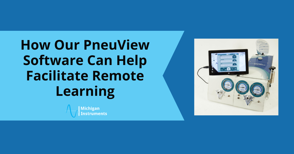
This PC-based software interfaces with our Lung Simulators, and accurately provides numeric and graphical display of ventilation parameters in real time. Additionally, it allows you to save and export data for later review and analysis.

These traits and more also make PneuView a great tool for remote learning. Keep reading to learn how.

1.   Real Time Displays

Volume, pressure and flow values and waveforms are displayed in real time. This makes it easy for educators to share their PneuView screen with students during remote learning sessions and explain each piece of data directly.

2.   Record and Playback Function

The PV3 software has a record and playback function. This way, you can run different scenarios and record them ahead of time to be played back during a remote learning session.

3.   All Information Displayed on One Screen

Some educators opt for the non-instrumented (non-PV3) versions of our devices, since many modern day ventilators are capable of displaying similar information and waveforms.

However, having this information on your computer instead, and all on one screen, easily facilitates sharing with remote learners!

4.   Easy to Learn and Use

With remote learning opportunities comes a sense of responsibility to learn and understand new teaching platforms and systems. PneuView is a valuable tool and is easy to learn and use.

It’s user-friendly for both educators and students, and informational while not being too complicated.

5.   We’re Here to Help

We have resources available to help guide the use of our PneuView software, so you can get the most out of your experience.

One of our resources is our YouTube channel. There are several videos available that  are valuable for understanding how PneuView works and taking advantage of all its benefits.

Additionally, our FAQ page contains a wealth of information about PneuView. It provides concise and easily accessible information that helps our customers find answers quickly, enhancing understanding and engagement of all of our products.

Finally, our tech support team is always happy to help with PneuView!

Learn More About Using PneuView to Facilitate Remote Learning

In the age of remote learning opportunities, discovering new helpful tools and devices can be the perfect solution for advanced education.

Using our PneuView software is a fantastic way to provide quality education alongside our lung devices. Contact us today to learn more!

asthma attack simulation

Asthma is a chronic condition that inflames and narrows the airways in the lungs. In fact, nearly 26 million people in the U.S. have asthma, making it one of the most common forms of respiratory disease in the country. 

Fortunately, you can easily replicate asthma attack simulation on a lung simulator for education and research purposes.

Why Use Michigan Instruments Lung Simulation for Learning about Asthma?

There are several reasons why our Lung Simulators are a fantastic tool for learning and teaching about asthma; including, but not limited to:

  • Safety: Simulation-based education ensures patient safety.  This allows the user to make mistakes and gives them the ability to correct them, without harm to a patient. 
  • Reduced Errors: Simulation provides opportunity for continued practice. This results in less error while working with humans.
  • Preparation for Several Scenarios: Simulation devices offer a wide range of lung compliance and airway resistance settings.  This offers the students and professionals an immersive and nearly hands-on experience. 

Settings For An Asthma Attack Simulation

By creating a severely restricted airway and pairing it with a slightly compliant lung, you can provide settings that simulate an acute asthma attack.

Here are the settings we recommend (using our lungs) to create a successful simulation:

  • Functional Residual Capacity: Should be between 860mL and 990mL per adult lung and 80mL to 200mL for the infant lung.
    • Please note that residual capacity is only considered functional if it’s downstream (during inspiration) of the airway resistor.
  • Airway Resistance: A resistance of 50cmH2O/L/s should be used to simulate this condition in adults. The user should use 500cmH2O/L/s to simulate this condition in infants.
  • Dynamic Compliance: Use a lung compliance of .04L/cmH2O for adults and .004L/cmH2O for infants. It’s important that this value is “dynamic” so that the compliance of the simulated lung will decrease as breaths begin to stack.

How is This Simulation Beneficial?

During normal ventilation, the increased airway resistance creates the high proximal pressure seen in victims of an acute asthma attack. 

Additionally, the above-average compliance of the simulated patient will cause breath stacking unless the breaths delivered are properly regulated.

You can test many simulations in a number of ways. This allows students to get hands-on experience with rare conditions, replicate case-specific failures in ventilators, identify pros and cons of different ventilation techniques and more.

Organizations around the world use our test lungs for a plethora of educational purposes. The flexibility of our equipment allows our devices to replicate hundreds of healthy and diseased lung conditions, like asthma, while providing accurate measurements and data that inform your testing, research, and training.

Learn More About Our Lung Simulation Capabilities

If there is a specific symptom or condition that you would like to replicate, please let us know. We’d love to put together a simulation for you.

For questions or more information about simulations, contact us today.

lung simulation model

Did you know that our lung simulator is used in many facilities that offer Respiratory Therapy programs—throughout the United States—and  around the world? 

Designed and manufactured by Michigan Instruments, our Lung Simulators are utilized in colleges, universities, hospitals and training schools. Their accurate and reliable human pulmonary system simulation capabilities make them an incredible tool for training and education.

Here are 10 reasons why we believe our equipment is a great solution for respiratory therapy programs.

Why Choose Our Lung Simulator for Respiratory Therapy Programs?

1. Lung Condition Simulation

Facilitators can simulate a wide variety of healthy and diseased lung conditions by altering the lung compliance and airway resistance in a one or two-lung simulation. 

The flexibility of the devices provide accurate measurements and data that inform testing, research, and training for any and all lungs!

2. Realistic Volumes

Students work with a realistic total lung volume and residual volume—especially when using the Dual Adult Lung Training & Test Lung

This is one of the many features that allows the devices to provide the best representation of the functions of the human pulmonary system.

3. Versatility

There is great versatility in regard to introducing gasses, tapping into the lungs or airways, and connecting auxiliary devices, like CO2 monitors.

This makes teaching easy and convenient, while providing a physical demonstration.

4. Ventilation Dynamics

With the Dual Adult Training & Test Lung, facilitators can simulate unilateral lung disease and the resulting ventilation dynamics.

This contributes to easy demonstration of mechanical ventilation and laboratory exercises with ventilators.

5. Spontaneous Breathing

You can simulate a spontaneous breathing patient and evaluate the response of devices in various support modes.

Spontaneous breathing simulations are especially helpful for designing, testing, and providing training on non-invasive and supportive modes of ventilation and oxygenation. Our SBL™ allows control of the breath rate, tidal volume, inspiratory time, and inspiratory flow pattern.

6. Multiple Techniques

Our SBL™ also facilitates education and studies with various oxygen delivery systems, and with the addition non-invasive ventilation devices & techniques. 

7. Helpful Displays

When you add the PneuView software, you are able to see real-time data and waveform displays of pressure, volume, and flow, even when using simple ventilation devices like bag-valve-mask (BVMs), emergency ventilators, and CPAP systems.

8. Recordings 

When using the PneuView software, facilitators and educators have the ability to capture ventilation data. This data can be graphed, tabulated, or digitally recorded and retrieved for later review, demonstration, and analysis. 

9. Save Data

Students should have a simple and convenient way to perform research and keep track of their findings—and our software is more than capable of achieving that. With our PneuView systems, students can conduct research and easily save their data.

10.  Easy and Durable

Last but not least, and probably the best benefit of our equipment in Respiratory Therapy Programs, is that it’s easy to use—and built to stand the test of time. 

Many facilities have been using our products for years.  The PneuView software was designed with simplicity in mind, and our software continues to evolve based on the needs of our customers. 

Our customers have been vocal about how our equipment has been able to benefit them:

“We have been using Michigan Instruments Training Lung Simulators in our Respiratory Care program for many years and we love them. The lung simulator easily provides simulation of a variety of lung conditions that students can adjust to complete a variety of lab exercises. They are versatile, and a great tool for giving students hands-on experience with mechanical ventilation techniques, simulation of disease states for understanding pathology, and much more!”

– Dr. Ann Flint, Program Director, Respiratory Care – Jackson College

Learn More About How Our Lung Simulators Benefit Respiratory Therapy and Other Educational Programs

Medical professionals, directors, and educators around the world have chosen to partner with us and use our equipment in their programs. Our Lung Simulation product line continues to expand  to better service Respiratory Therapy Programs everywhere.

If you’re interested in learning more about our lung simulation devices, the PneuView Software, or any of our additional products, contact us anytime for more information.

importance of continuing education ems

This year, May 21st – May 27th is National EMS Week. This week is dedicated to celebrating EMS employees and the important part they play in our nation’s communities to help save lives.

In light of this annual event, we would like to acknowledge the importance of proper education and technology for your EMTs. 

Below are the different ways to provide your EMTs with continuous education, and all the benefits that they provide. 

1. Understand Why Continuing Education is Important

All EMS organizations should offer continuing education within their facility, or in partnership with other healthcare organizations. However, continuing education for EMTs offers more benefits than just filling a requirement to maintain certifications. 

There are several benefits to providing your EMTs with education indefinitely, such as:

More Effective and Efficient Treatment

Keeping up with the latest medical information and technology allows EMS teams to provide better treatment for their patients. 

New practices and technologies (like ours) can save emergency medical professionals time by treating patients with methods that may not have been previously available.

Reduced Risk

Continuing education also reduces liability risk from malpractice or wrongful death. Failure to be consistent with education can result in incorrect treatment, misdiagnosis, or other life-threatening mistakes.

Networking Opportunities

In-person education programs, such as classes and conferences, allow EMS administrators to build connections and grow their network. Forming relationships in the industry can open new career paths, and create opportunities for shared knowledge.

2. Explain the Importance of Learning from Others

No matter what role you play or how long you may have been performing your job,  you should never assume there isn’t more to learn—especially in healthcare. Healthcare professionals should always be willing to learn new information or gain new perspectives from others. 

This also includes helping them acknowledge the value of feedback, and doing self-assessments to better understand their strengths and weaknesses.

3. Be Open to Multiple Teaching Techniques

While all of your EMTs should remain open to learning new things, they might not have the same preferred learning styles or methods. 

Some learn best in a classroom-like environment, while others may prefer educational apps, medical devices, academic journals, or mentors. Keep your options open when looking for educational opportunities for your staff.

The National Association of EMS Educators offers many options for educating EMS Professionals. This is just one of the many ways to find resources that you can use to craft an educational plan for your EMTs.

4. Select Your Best Personnel for QI Leadership

In order to have an effective quality improvement (QI) program, it is understood that providers want to do a better job of providing patient care. If your QI program is successful, it will help your EMTs recognize opportunities within their patient care and encourage them to make proper corrections.

If your QI program is less successful, you may have the wrong leader and might consider making a change to ensure more effective leadership.  

Enlist the help of your best and most qualified clinicians to do QI. Teach them about good culture, statistical process control, normal and special cause variation, and other improvement science concepts that drive them to improve care and be better leaders for the team.

5. Commit to Communication

One of the key reasons why things can go wrong in healthcare organizations is a lack of communication.  There is a failure to communicate issues, questions or changes that would benefit the team. 

When discovering concerns and/or opportunities to improve, it’s vital to share them with your staff. Encourage your team to practice continuous communication no matter what. 

An operation without communication is a recipe for disaster. In an environment where everyone is working collaboratively to save lives, all aspects of communication—including addressing errors, offering constructive feedback, and updating staff on changes in practices—are of utmost importance for a successful operation.

6. Provide Your Team With Michigan Instruments’ Medical Devices

Medical technology has been known to be an avenue for effective education for many healthcare professionals—especially those who practice emergency services. 

For example, our lung simulators can provide helpful training and practice for EMTs. They are fully adjustable and provide a dynamic response to therapy that accurately demonstrates how the human pulmonary system works. 

In addition, we offer outstanding medical equipment, such as our automated CPR machines. These help EMS teams to be more efficient and effective.  

Our Life-Stat and Thumper CPR devices support a quick and simple transition from manual to automated CPR with minimal interruption to compressions. 

The Hands-Free operation of our devices and the option to automatically ventilate the patient along with chest compressions gives the EMS team more time to focus on other life-saving activities. 

While very cost effective and easy to operate, these devices also can perform CPR on larger patients compared to other competing devices.

Learn More About How Our Devices Benefit Emergency Medical Practices

The importance of continuing education for your EMS team cannot be emphasized enough. However, our devices can grant your team new opportunities for both continuing education and effective patient care. 

In honor of National EMS Week, contact us today for more information about why thousands of medical professionals all over the world choose Michigan Instruments.

recruit medical students

As the healthcare industry continues to grow, so does the demand for qualified medical professionals. To do this, you need to recruit medical students to your program.

However, with so many accredited medical programs available, it’s now more difficult than ever to attract students to your program. To help you stand out among other programs, keep reading to learn about ways to attract students to your medical program.

How to Recruit Medical Students to Your Program

1. Emphasize Your Program’s Unique Features

It’s important to highlight the specific aspects of your program that set it apart from others. This could include specialized training in a certain area, access to state-of-the-art technology, or opportunities for hands-on experience.

Students want to know what to expect when coming into a new program, so be transparent and explain to them all the benefits you want them to know about. This way, they can make an informed decision when they’re choosing between programs.

2. Share Success Stories

Prospective students want to know that your program is going to help them achieve their career goals. To demonstrate the knowledge and skills that your program can give them, share stories of alumni who have gone on to find successful careers in the medical field and highlight how your program helped them get there.

This will not only show your capability of teaching students the curriculum they need, but will also help them find inspiration in others that have done the same program. Remember, word of mouth is a powerful tool.

3. Use Social Media

Social media platforms, like TikTok, give you the opportunity to showcase your program’s offerings and connect with potential new students. Share photos and videos of your facilities, faculty, and students to give a behind-the-scenes look at your program.

It’s important to be informative—however, it’s just as important to craft personal messages to connect with your audience.

4. Offer More Scholarships and Financial Aid 

The cost of medical programs can be a major barrier for a lot of students. Offering scholarships and financial aid can help attract students who may not have otherwise considered your program.

There are lots of resources available for more information about providing financial help. Before doing so however, you need to read up on the IRS requirements for starting a scholarship fund.

5. Utilize Advanced Technology

Medical technology has made impressive advances in the past decade. For instance, our lung simulators can provide helpful training and practice for medical students, particularly in the Pulmonology and Respiratory fields.

Our Lungs offer residual lung volumes and a dynamic response to therapy that realistically demonstrates how the human pulmonary system works. 

What’s the Difference Between Michigan Instruments’ Lung Simulators and Other Devices on the Market?

Unlike other devices on the market today, our lung simulators are available in both adult and infant models. This range of calibrated lung compliance and airway resistance settings allow our devices to replicate hundreds of healthy and diseased lung conditions, while providing accurate data to contribute to your testing, research, and training.

Our recently launched Spontaneous Breathing Lung is a great addition to any respiratory therapy program. The SBL™ provides accurate simulation of a breathing patient and is available on any of our lung simulators. It’s beneficial for designing, testing, and training for non-invasive and supportive modes of ventilation and oxygenation. 

With our SBL™, the sky’s the limit. Our devices have been used for almost everything—from studying the dispersion of aerosols and inhalers, to assessing the responsiveness of the latest technologies. 

Learn More About How Our Devices Can Benefit Your Medical Program

For more information about our devices and how they can better your program, contact us today and get in touch with a service specialist that can answer all your questions!

michigan lung simulator

For over 45 years, our Michigan Lung Simulator has offered residual lung volumes and a dynamic response to therapy that realistically represents all the functions of the human pulmonary system. Our devices are more advanced now than ever, thanks to our ever-changing technology and dedication to research.

We receive many questions about our lung simulators and their operations. Below are the questions we see most often

1. What Is The Difference Between A “Test Lung” And A “Michigan Lung Simulator”?

We’ve used the terms “test lung,” “training test lung,” and “Michigan lung simulator” to describe our TTL® and PneuView® products. In some ways, these terms are interchangeable. However, in a broader sense, the term “test lung” may include devices that are very simplistic rubber or latex bags. 

On the other hand, “lung simulators” describe a more complex system that accurately mimics the dynamic mechanical characteristics of the human pulmonary system.

2. What Is The Fundamental Purpose Of A Michigan Lung Simulator?

There are several situations and settings where the use of a lung simulator is crucial. A solid lung simulator represents the functions of the human pulmonary system, as well as a range of healthy and diseased lung conditions. 

A lung simulator should allow you to create, monitor and control those forces.  For the following applications and more, a high-quality lung simulator is needed:

  • Designing mechanical ventilators and other respiratory apparatus
  • Developing new modes of ventilation support
  • Training respiratory care and other medical professionals
  • Performing periodic testing and maintenance on ventilation and support devices
  • Troubleshooting equipment problems using a realistic “load”

3. How Do The TTL® And PneuView® Systems Model The Dynamic Compliance And Resistance Characteristic Of The Human Lungs?

Our TTL® and PneuView® systems use a bellows and spring to simulate the compliance characteristics of the lung. The spring can be positioned at several different points along the Top Plate of the simulator to create a lung compliance that ranges from very compliant to normal to very non-compliant (or stiff). 

The resistance characteristics of the lung are set using fixed-orifice parabolic resistors that may be positioned to add resistance to the upper and/or lower airway assembly.  A range of resistors allow for simulation of both healthy and diseased lung conditions.

4. What Is Factory Calibration?

Factory calibration involves tuning all aspects of sensor, electronic, mechanical and software to ensure maximum accuracy of the measurements and simulated parameters provided by TTL® & PneuView® systems. 

Calibration procedures include the setting of the compliance and resistance characteristics of the lungs, as well as setting offset and gain characteristics for each of the pressure transducer channels.

5. How Do I Read The Tidal Volume On The Lung Simulators?

Each simulated lung in the TTL® or PneuView® System has a physical volume scale behind the Top Plate. There is also a plastic pointer and label on the Top Plate that can be used to indicate lung volume. 

To ensure the most accurate indication of lung volume, the pointer on the top plate must correspond to the compliance setting on the lung. When set appropriately, the arrow will point to the gas volume in the lung. 

Note: When using PEEP or CPAP, the starting volume (baseline volume) is greater than zero. The tidal volume would be the total indicated volume minus the baseline volume.

The volume indicator lines on the label are curved, more so as the compliance setting decreases. This is due to the sideways distention of the bellows that occurs during filling of the lung under pressure.

6. Are Michigan Lung Simulators Suitable For Tests With Aerosols Or Just Dry Air Only?

Water vapor will not damage the simulator.  However, we generally don’t recommend introducing aerosolized substances into the lung chambers of our TTL® and PneuView® simulators for the following reasons:

  • Substances other than water can be corrosive to the polyurethane bellows, and/or may accumulate on the ribs of the bellows causing them to stick, become brittle, or not function properly.
  • There is no simple way to “drain” the bellows of any accumulated fluid. If using sterile/demineralized water, you may blow dry gas through the unit until it is fully dried.

Many of our customers have used the TTL® or PneuView® specifically to evaluate aerosol delivery with various devices and/or breathing patterns. In such cases, the aerosolized substance is typically collected using a hydrostatic filter placed in the simulated airway, prior to entering the lung bellows.

Let Us Answer Your Questions

If you have any other questions about our lung simulators, visit our FAQ page or contact us directly!

Lung Simulators: Why Choose Michigan Instruments?

Lung simulators are designed to replicate the function of healthy and diseased human lungs for testing, teaching and training. Comparing the features, settings and the realistic nature of a lung simulator is vital to its success and effectiveness.

Below, we outline the 3 main features of our lung simulators.

1. Realistic Appearance and Feel

When utilizing lung simulators in teaching, training and testing, it’s important to create the most realistic scenario as possible.  

Michigan Instruments’ simulators do that as they are built fully to scale, and offer realistic residual lung volumes and capacities. Our devices move, breathe and feel like real lungs when ventilated. 

Additionally, our Training Test Lungs (TTL) and Pneuview Systems are available in adult and infant models. 

2. Enhanced Lung Simulator Settings 

When using our devices, it’s best to have a varied range of settings for compliance and resistance. Our TLL and Pneuview systems provide enhanced settings that exceed the capabilities of many other simulators on today’s market.

Our devices include a variety of settings and capabilities, such as:

  • different lung conditions and pathologies,
  • tools for teaching and training, and 
  • measurements for lung volume, lung and airway pressure, and more.

3. Affordable Cost

Michigan Instruments’ lung simulators and PneuView systems offer your institution a realistic simulation, with an array of features and settings—at a fraction of the cost of other devices available.

Which One is Best For You?

While both the TTL and Pneuview Systems offer a variety of dynamic features and simulations, it all comes down to which one is best for your organization’s training, testing and research needs.

Learn more about our high-quality lung simulators that you can touch, see, and modify and the differences between each one. Questions? Request a quote, or ask us anything!